Green Gram Cowpea Weevil

(Callosobruchus maculatus)

Credits: Biovision-Infonet

The cowpea weevil (Callosobruchus maculatus) on cowpea (Vigna unguiculata) seeds. Adults are 2 to 3.5 mm long
(c) Peter Credland. Reproduced from the Crop Protection Compendium, 2006 Edition.
The larvae and pupae of the cowpea weevil Callosobruchus maculatus) are normally found in the seeds of pulses. Upon hatching, the larva bites through the base of the egg and bores into the seed, where it spends the whole lifecycle feeding on the seed. The larva pupates inside the seed.
(c) F. Haas, icipe

Cowpea bruchids (Callosobruchus spp.) are the most common and widespread insect pests in storage. Adults are 2 to 3.5 mm long. They are major pests of pulses (cowpeas, pigeon peas, soybean, green gram and lentils). They attack both pods in the field and seeds in storage. They attack nearly mature and dry pods. Infested stored seeds can be recognised by the round exit holes and the white eggs on the seed surface. Post-harvest losses are highly variable, but losses can be over 90%.

It is a serious storage insect, which can destroy whole seed-lots.

What to do:

  • Dry grains to moisture level below 13%.
  • Store grains in dry, well ventilated areas.

Green Gram Production

Credit: Biovision-Infonet

Green gram seeds
(c) Courtesy EcoPort Pankaj Oudhia
Mungbean plant
(c) A.A.Seif, icipe
Mungbean crop
 (c) A.A.Seif, icipe

Scientific Name: Vigna radiata

Order / Family: Fabales: Fabaceae

Local Names: Pojo (Swahili)Common Names: Mungbean

Pests & Diseases: Anthracnose, Aphids, Bacterial blight, Bean flies, Cowpea seed beetle, Damping-off diseases, Foliage beetles, Mungbean Yellow mosaic virus, Pod sucking bugs , Pod-borers, Powdery mildew, Rust, Storage pests, Thrips, White

Geographical Distribution in Africa

Geographical Distribution of Green gram in Africa. Updated on 8 July 2019. Source PROTA

General Information and Agronomic Aspects

Grams are annual legume crops grown for their seed. Grams could be green, black or yellow in colour. The green grams are the most commonly grown in Kenya. Grams are native crops of India. Often called green gram or golden, it is cultivated in several countries of Asia, Africa, and the Americas. The dried beans are prepared by cooking or milling. They are eaten whole or split. The seeds or the flour may be used in a variety of dishes like soups, porridge, snacks, bread, noodles and even ice cream. Green gram also produces great sprouts, which can be sold in health food shops or eaten at home. Crop residues of V. radiata are a useful fodder. Green gram is sometimes specifically grown for hay, green manure or as a cover crop.

Climatic conditions, soil and water management

Green grams grow best at an altitude of 0-1600 m above sea level and under warm climatic conditions (28 to 30degC). They are well adapted to red sandy loam soils, but also do reasonably well on not too exhausted sandy soils. Green grams are not tolerant to wet, poorly drained soils. They are drought tolerant and will give reasonable yields with as little as 650 mm of yearly rainfall. Heavy rainfall results in increased vegetative growth with reduced pod setting and development. 

Propagation and planting

Avoid planting green gram for more than one season because toxic residues and disease organisms from the previous green gram crop may affect the following crop adversely.

 Land should be prepared to a medium tilth before planting and early enough so that planting can start immediately after the rain starts. When using oxen plough for planting, place the seed at the side of the furrow.

 Propagation is by seed. There is no seed dormancy. Seeds may sprout in the pod under very humid conditions. In areas with higher rainfall, it is recommended to grow green grams on raised beds. Prepare the beds, raised about 20 cm and spaced 1 m from the centre of one bed to the centre of the next. Sow seeds on raised beds in two rows per bed, spaced 45 cm apart. 

Green grams will respond to fertiliser or manure application but will normally give satisfactory results if grown on relatively good soil. Green gram is grown mainly on smallholdings, often as mixed crops or intercrops. Associated crops are usually of longer duration than green gram (sugar-cane, cotton, sorghum). To make use of a short cropping period, short-duration green gram is often relay-cropped.  

Green gram (mung beans) varieties

Mungbean plant
(c) A.A.Seif, icipe

Mungbean crop
 (c) A.A.Seif, icipe
Green gram seeds
(c) A.A.Seif, icipe

Green grams usually mature in 60 to 90 days. The early maturing varieties can often produce before drought destroys many bean species. Two varieties can be distinguished in Kenya:

VarietyMaturity DaysPotential yield t/haRemarks
“KVR 22” (“N 22”)80-901.0-1.3Golden yellow seed colourTolerant to aphidsResistant to yellow mosaicModerately resistant to powdery mildewIn the driest areas will perform poorly due to its latenessPerforms well between 50 and 1600 m above sea level
“KVR 26” (N 26)60-650.3-1.5Shiny green seed colourBest performer in dry areas due to its earlinessPerforms well between 50 and 1600 m above sea level

Husbandry

With the newer cultivars ripening in 60 to 75 days, maximum yields are obtained at plant densities of 300 to 400,000 plants per ha. The later-maturing traditional cultivars generally need wider spacing. Usually no fertilisers are applied to green gram. Over the centuries, green grams adaptation to stable performance in marginal environments has resulted in a low yield potential, which limits responsiveness to better environments and improved cultural practices. However, if planted in heavily eroded soil gram will benefit from any kind of manure or compost. 

Grams planted at the end of the long rains are normally intercropped into other major crop. In Meru, Kenya, green gram is a preferred intercrop for millet, each said to protect the other against diseases and pests. If grams are intercropped with maize, the maize spacing is the same as in pure stand, but the grams are interplanted mid-way between the maize rows. Early weeding is recommended. First weeding should be done just after emergence and second weeding just before flowering.  

Harvesting

Harvesting is generally by two to five hand-pickings at weekly intervals and is the most expensive single operation in growing green gram. Short-duration cultivars, which ripen more uniformly, may be processed as whole plants on small rice threshers.Cultivars differ markedly in harvesting efficiency, depending on position (above or within canopy) and size of pods. Harvesting before the maturity of crop, usually result in lower yields, higher proportion of immature seeds, poor grain quality and more chances of infestation during storage. Delay in harvesting results in shattering of pods and other losses caused by pests.

In Kenya, harvesting when 95% of pods have turned black is recommended. The whole plant can then be uprooted and dried for about 2 days, then threshed and winnowed. Harvesting during adverse weather condition i.e. rains and overcast weather should be avoided. Such weather is conducive to fungal infection. The harvested bundles should be kept in one direction in order to ascertain efficient threshing. They should be stacked in a dry, clean place in cubical way to facilitate circulation of the air around. 

 Storage

Grams must be dry before storage. Like most pulses moisture content at storage should not be above 13%. Grams are very susceptible to bruchid (bean weevil) attack and are best stored immediately after sun drying either in airtight drums tins, gunny bags and be kept in a clean, ventilated place. Mixing seed with ash is effective against bruchids, also treatment with sunflower oil or mixing with neem leaves is said to be effective against storage pests. Proper drying of grains is very important to prevent the growth of fungi and contamination with aflatoxins. Infected grains should be separated from sound grains to avoid aflatoxin contamination.

Q fever

Q fever description

Q fever is a bacterial disease that affects both humans and animals. It is caused by Coxiella burnetii

Q fever has a worldwide distribution with exception of New Zealand

Q fever animal Symptoms

The most common sign of infection in animals is abortion during late pregnancy. 

Most animals do not show any signs of illness with Q fever including lack of fever!

Q fever Transmission in animals

The bacteria mainly affects farm animals like cattle, sheep and goats. They then infect a variety of other species like humans, rodents, pets, birds, and fish

Direct contact is the main route of infection in animals through inhalation of contaminated air as well as contaminated farm equipment especially during birth

Ticks play a major role of maintaining the infection between animals and therefore importance of proper tick control

Dogs may be infected through consumption of placentas or milk from infected ruminants, and by the aerosol (air) routes. 

Q fever human Symptoms

Symptoms of Q fever include fever, chills, night sweats, headache, fatigue and chest pains. 

Pneumonia (lung infection) and hepatitis (inflammation of the liver) can occur in serious cases. 

In pregnant women, infections can cause premature delivery, abortion and infection of the placenta. 

In people with pre-existing heart valve disease, endocarditis (inflammation of the heart valves) may occur.

In pregnant women, Q fever can lead to spontaneous abortion, premature births and death in utero

Q fever Transmission in humans

Human cases are often associated with proximity to ruminants particularly at birth or during abortions and dry, windy weather

Humans get infected by breathing contaminated dust, assisting with delivery of newborn animals, contact with infected wool, urine, faeces, and tick bites

Sick animals also shed bacteria in milk, and thus, consumption of raw milk or their products e.g. yoghurt, cheese could be a source of infection to humans.

Dairy cows are more frequently infected for a long time and thus represent the most important source of human infection 

Q fever prevention

Keep pregnant livestock separate from other animals.

Burn or bury the remaining reproductive tissues after abortions or delivery of newborn animals to reduce the spread of the disease between animals.

Take great care when handling animal tissues to avoid your exposure to Q fever. 

Pregnant women should avoid contact with domestic animals or pets, to prevent infections with Q fever

Avoid contact with animals and birth materials, especially while animals are giving birth. Animals can be infected with Q fever, but may appear healthy

Eat and drink only pasteurized milk and milk products.

If you are assisting the delivery of newborn animals, wear gloves, masks and eye protection

People with heart valve disease, who have had valve replacements or pregnant women should be especially careful around pregnant sheep, cattle and goats.

Q fever Action

If you suspect Q fever contact your veterinarian for information on how properly to dispose of possibly infected tissue.

If you suspect Q fever: (1) In animals – contact your veterinarian; (2) In humans – contact your physician 

Content provided by Zoonotic and Emerging Diseases Group (ZED) University of Liverpool, International Livestock Research Institute (ILRI), Zoonoses in Livestock in Kenya (ZooLink)

Rabies

Rabied Dogs
Photo Credit ILRI/ZooLink

Rabies Description

Rabies is a fatal viral disease affecting all mammals and most often transmitted to humans from rabid animals through bites or scratches

In 99% of cases, domestic dogs are responsible for rabies virus transmission to humans. The disease is almost always fatal following onset of clinical signs

Rabies is present on all continents, except Antarctica, with over 95% of human deaths occurring in the Asia and Africa regions.

In many developing countries (including Kenya), unvaccinated dogs are the primary source of infection for humans and almost always ends in death. 

Cow with Rabies Photo Credit ILRI/ZooLink

Rabies animal Symptoms

All mammals can get rabies. Most cases in domestic animals species occur in cats, dogs, cattle, and horses. In wildlife, bats can be a source of infection.

Signs of disease involve changes in behaviour, such as a friendly dog that becomes withdrawn or aggressive, or lack of fear in cattle

Other signs include restlessness, paralysis of their legs, difficulty swallowing, a lot of salivation and a change in the sound of their voice. 

Affected animals overreact to stimuli such as noise or light. 

Any animal infected with rabies will die in 7 to 10 days.  

Rabies Transmission in animals

Rabies is most commonly spread among animals by the bite of an infected animal whose saliva contains the virus. 

Open wounds, the eyes, nose, and mouth are also possible ways of becoming infected when in contact with saliva from an infected animal.

The virus is not thought to be spread by blood, urine or faeces.


Rabies human Symptoms

In most human cases, signs of rabies develop 1 to 3 months after exposure.

Early symptoms include fever, headache, itching at the site of the bite, confusion and abnormal behavior.

Hypersensitivity to light and sound, and difficulty swallowing can also occur. 

Once signs of disease begin, recovery is very rare. Death usually occurs within 2 to 10 days. 

Treatment before signs develop is highly effective and life-saving. 

Rabies Transmission in human

Humans are usually infected following a deep bite or scratch from an animal with rabies. 

Transmission can also occur when saliva from an infected animal comes into direct contact with human eyes, mouth, nose or fresh skin wounds

Other minor/rare routes are contamination of the eyes, nose and mouth, air transmission and organ transplantation

Rabies prevention

Animals should be vaccinated for rabies and kept away from wildlife. 

Dogs, cats or ferrets that have bitten humans and show no signs of illness may be observed for rabies under veterinary supervision for 10 days. 

Do not contact, handle or feed wild animals and report stray animals to your veterinarian.

Wildlife or domestic animals with unusual behavior should especially be avoided. 

Do not pick up bats, dogs, cats or any animal that are laying on the ground. Report them to your nearest veterinarian immediately. 

If you are bitten by any animal, wash the wound well with soap and hot water and contact your physician immediately.

Create awareness on dog behavior and bite prevention among children and adults and the importance of rabies vaccination

Rabies Action

Prevention is the best way to stop rabies. Therefore ensure that your animals (especially your dog and cat) are vaccinated against rabies

If you suspect rabies: (1) In animals – contact your veterinarian; (2) In humans contact your physician 

Content provided by Zoonotic and Emerging Diseases Group (ZED) University of Liverpool, International Livestock Research Institute (ILRI), Zoonoses in Livestock in Kenya (ZooLink)

Trypanosomiasis

Cow with Trypanosomiasis
Photo Credit ILRI/ZooLink

Trypanosomiasis description

Trypanosomiasis is a disease of humans and animals caused by blood parasites and transmitted by tsetse flies.

Trypanosomiasis is called Nagana in animals and sleeping sickness in humans. 

Nagana causes serious economic losses in livestock from anemia, loss of condition and emaciation. Many untreated cases are fatal

In humans, sleeping sickness is commonly found in 37 African countries geographically distributed in two forms and fatal if not treated. 

One form is endemic in 24 countries West and Central Africa while the other form is endemic in 13 countries of Eastern and Southern Africa 

Transmission of Trypanosomiasis

The parasite develops in the tsetse fly and is transmitted into animals/ people through a bite. The parasite develops in the animal/ humans causing sickness. 

Trypanosomes can also be spread by equipment we use on farm such as surgical instruments, needles, syringes and various biting flies including horse flies 

Trypanosomiasis animal symptoms

The disease can affect cattle, water buffalo, sheep, goats, camels, horses, donkeys, pigs, dogs, and cats

Symptoms of disease in animals will occur as from 4 days to approximately 8 weeks after infection 

The main signs of trypanosomiasis in animals are: fever, swollen lymph nodes, weight loss, anaemia, decreased milk production and abortions.

Trypanosomiasis human symptoms

Symptoms in humans occur in two stages: (1) Early stage and (2) Late stage

Early stage symptoms are non-specific and often un/misdiagnosed e.g. fever, irritability, fatigue, swollen glands, aching muscles and joints, and headaches

Late stage symptoms cause serious mental disorders including sleeping problems and mental problems 

If trypanosomiasis is untreated in people, infection becomes worse and death occurs within months.

Trypanosomiasis prevention and control

Nagana can be managed by reducing tsetse fly populations, breeding trypanotolerant animals and treating infected animals with antiparasitic drugs

Vector control for trypanosomiasis includes clearing vegetation and bushes, grazing away from tsetse-infested areas, insecticide ground and aerial spraying 

Drugs that kill trypanosomes are available and are used in the treatment of sick animals.  

In humans, prevention of trypanosomiasis is avoiding tsetse fly bites, by wearing protective clothing, using insect repellant, avoiding bushes in infested areas

Treatment of trypanosomiasis in people requires a diagnosis of infection by a medical practitioner before drugs are given.

Warning for Trypanosomiasis 

If you suspect your animals have Trypanosomiasis contact your veterinarian immediately to work on a management plan

In humans, if one suspects they have Trypanosomiasis, they should consult a medical health care provider for confirmation and treatment.

Content provided by Zoonotic and Emerging Diseases Group (ZED) University of Liverpool , International Livestock Research Institute (ILRI), Zoonoses in Livestock in Kenya (ZooLink)

Rift Valley Fever

Aborted foetus
Photo Credit ILRI/ZooLink

Rift Valley Fever Description

Rift Valley Fever or RVF is an acute fever-causing disease affecting animals (cattle, buffalo, sheep, goats, camels) that can also cause illness in humans.

Rift Valley Fever Virus is mainly transmitted by mosquitoes, so outbreaks are mostly seen during heavy rainfall seasons.

Rift Valley fever occurs throughout most of Africa, and has been reported in Kenya.

Rift Valley Fever Symptoms in animals

Sheep, cattle, and goats are the most severely affected. Other animals such as water buffalo, camels, rodents, cats, and dogs can also be infected. 

Rift Valley fever tends to affect young animals more severely than mature animals.

In young animals, signs of infection include fever, failure to eat, weakness, diarrhea and sudden death with no major signs of illness.

In older animals, infection may cause fever, discharge from nose, weakness, diarrhea, vomiting, decreased milk production and abortion. 

Abortion is often the only sign in mature animals.

Some RVF affected animals may have bloody diarrhea; salivation; nasal discharge and some may have yellowing of the eyes and mouth (jaundice) 

During outbreaks of RVF there are increased cases of newborn deaths, abortion and fetal malformation. The aborted fetuses may look digested 

Rift Valley Fever transmission in animals

Animals get RVF from infected mosquito bites; animal-to-animal infection can also happen through direct contact with infected tissues/fluids

Infection with RVF might also happen during the re-use of needles during treatment/ vaccination especially when veterinary resources are limited

Rift Valley fever can also possibly be spread by the bite of infected ticks and biting midges.

Rift Valley Fever symptoms in humans

The disease might not have any symptoms or may present as acute fever with headaches and weakness for 10-14 days before recovery 

Severe cases of RVF may lead to liver failure; kidney failure; vomiting blood; bloody stool, or bleeding from gums, skin, nose, and injection sites

Rift Valley Fever transmission in humans

Humans can get RVF from bites of infected mosquitoes but are mainly infected after exposure to blood, body fluids or tissues of infected animals.

RVF exposure happens mainly with people working with animals/their carcasses e.g. butchers, slaughterhouse workers and veterinarians.

Severe RVF infection occurs during outbreaks; no human-to-human transmission has been recorded

Death can occur in rare cases and is more likely when the severe form of the disease develops.

Rift Valley Fever Prevention

To prevent RVF infection avoid direct contact with animal blood, body fluids or tissues of infected animals

Avoid slaughtering of animals in areas suspected to have an RVF outbreak to avoid contact with infected livestock

To prevent RVF infection control mosquitoes especially in prolonged rainy seasons, use mosquito repellents and bednets.  

Animals in high-risk areas should be vaccinated to protect them against RVF infection

Raise awareness and educate friends and family about signs, symptoms and risk practices associated with RVF infection

Rift Valley Fever Warning

RVF is a notifiable disease; therefore report any suspected case to veterinary authorities in your area

If you suspect RVF, DO NOT touch the aborted fetuses/ blood/ body fluids/ tissues of infected animals!

To prevent RVF infection do not slaughter sick animals or eat meat from affected animal during an outbreak

If you suspect RVF: (1) In animals – contact your veterinarian; (2) In humans – contact your physician 

Content provided by Zoonotic and Emerging Diseases Group (ZED) University of Liverpool , International Livestock Research Institute (ILRI), Zoonoses in Livestock in Kenya (ZooLink)

Leptospirosis

Leptospirosis Description 

Leptospirosis is caused by spiral shaped germs (bacteria) called Leptospira that damage the liver, kidneys and other organs of animals and humans

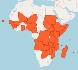
Leptospirosis as a disease occurs worldwide; with outbreaks mainly occurring after floods

Leptospira, are excreted through urine of infected animals into the environment where it survives for weeks and months.

Leptospirosis Transmission in humans

Agricultural workers and those working with livestock or wild animals are most at risk of leptospirosis. 

Leptospira bacteria gets into the body through broken skin (from a cut or scratch) or through the eyes, nose or mouth.

Contact with urine from an infected animal or urine contaminated soil, water or food exposes humans to infection and can survive there for weeks to months

Contact with other body fluids (except saliva), tissues and organs from leptospirosis infected animals can also cause human infection.

Exposure to leptospira contaminated waters i.e. flood waters and rivers cause leptospirosis outbreaks with person to person transmission being rare.

Leptospirosis Symptoms in humans

Leptospirosis in humans present with non-specific symptoms hence can be mistaken for other diseases 

Leptospirosis symptoms range from none, mild to severe often due to lack of treatment and death in rare cases.

Mild leptospirosis present with; high fever, headaches, chills, muscle aches, abdominal pain, diarrhea, vomiting, yellowing of the skin and eyes and red eyes.

Severe leptospirosis present with kidney and/or liver damage, infection of the brain and spinal cord, respiratory distress and even death.

Leptospirosis Transmission in animals 

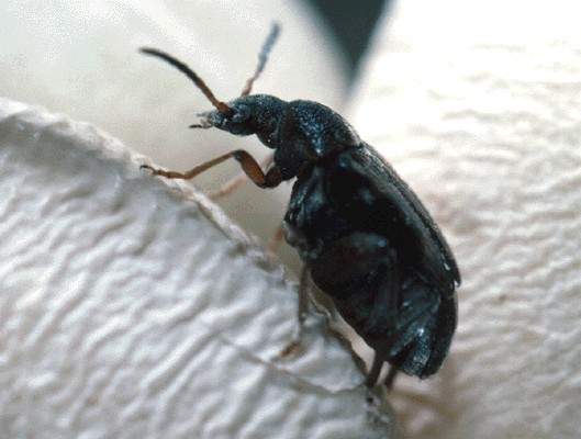
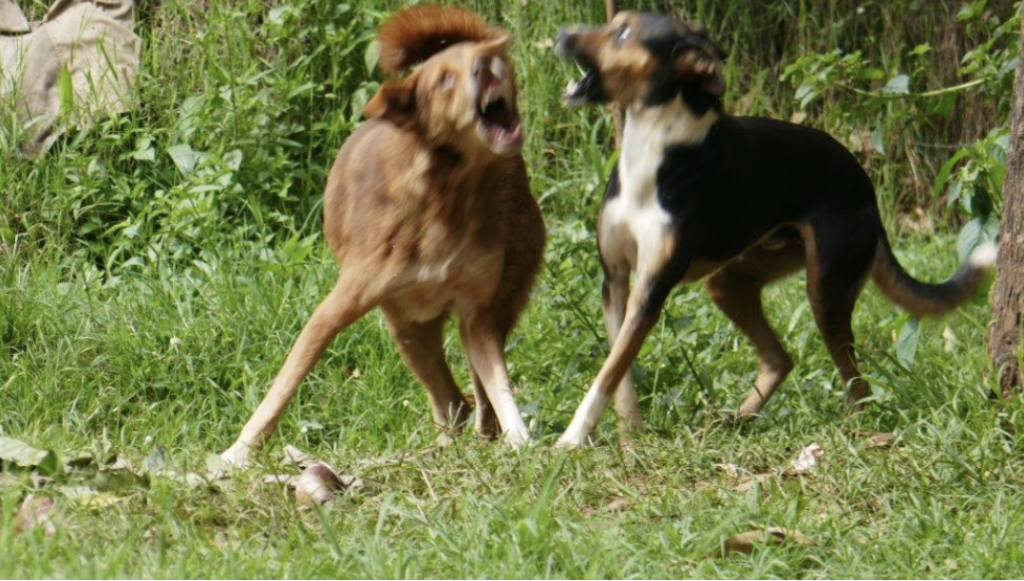
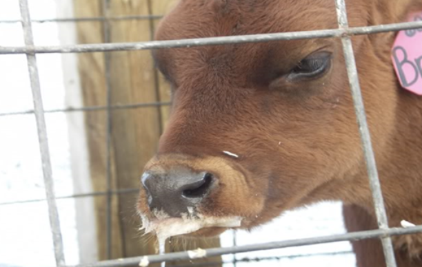
Leptospirosis affects cattle, pigs, sheep, goats, rabbits, horses, dogs and rodents.

Leptospirosis is spread through the urine of infected animals, which gets into water or soil and can survive there for weeks to months.

Animals become infected when they come into direct contact with this contaminated water or soil. 

The bacteria enter through cuts in the skin or through their eyes, nose or mouth.

Animals can get leptospirosis if their eyes, nose, mouth or broken skin come into contact with infected urine, contaminated soil, water, feed, or bedding

Contact with other unvaccinated livestock and/or rotational grazing may also expose livestock to leptospirosis.

Leptospirosis can also be spread from mother to offspring through the placenta, sometimes through breeding and from a bite by an infected animal.

Eating infected tissues can also expose animals (i.e. dogs) to leptospirosis

Leptospirosis Symptoms in animals 

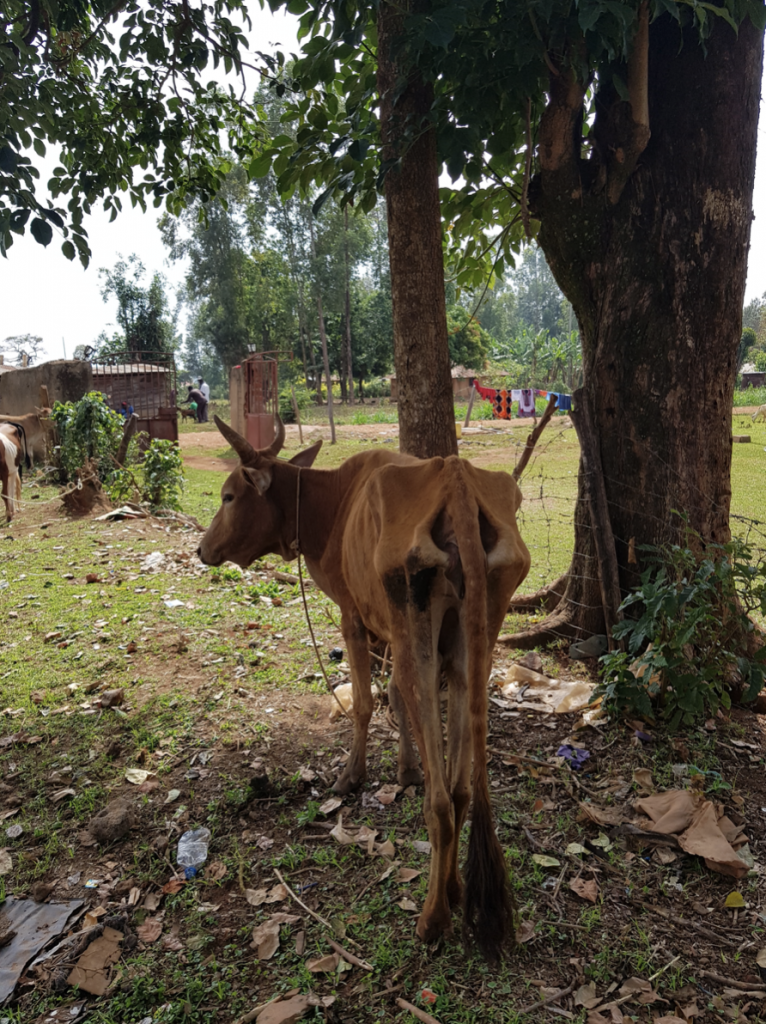
Many animals do not show any signs of illness but they continuously contaminate feed, water, and pastures with the germ causing leptospirosis through urine

General symptoms include: fever, shivering, muscle pain, decreased activity, increased thirst, vomiting, diarrhoea and changes in frequency of urination

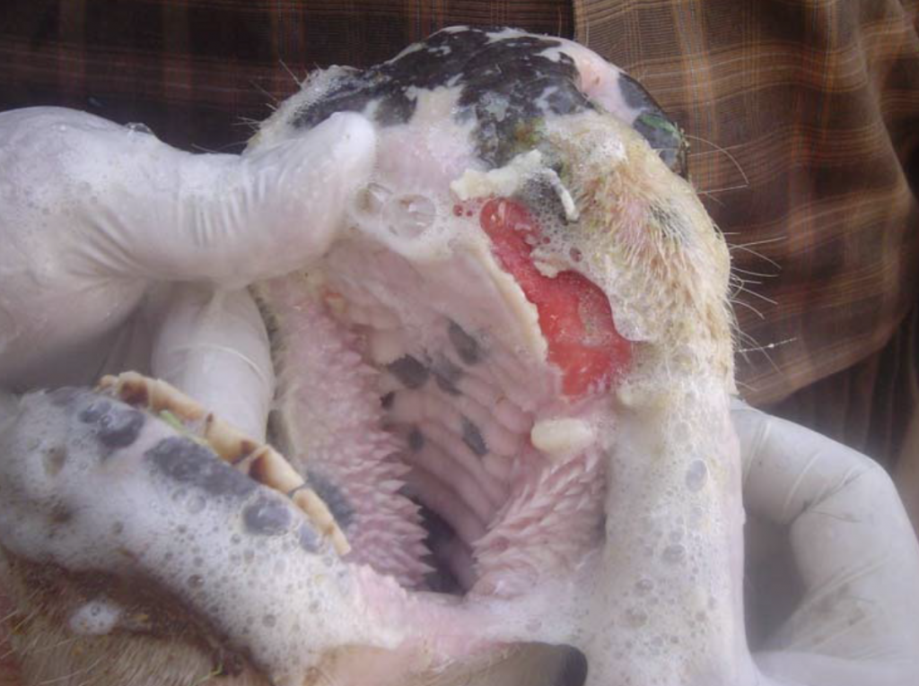
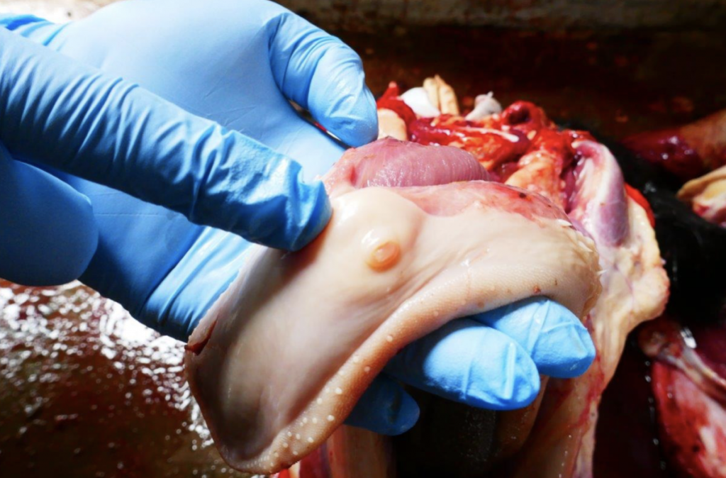
Leptospirosis causes bleeding disorders with blood-tinged vomit, urine, stool or saliva; nosebleeds and red spots on gums, other mucous membranes or on skin.

Some animals also develop swollen legs or accumulate excess fluid in their chest or abdomen.

Lactating cows and sheep will have mastitis causing the udder to suddenly become sluggish with resultant milk drop and changes in color and consistency of milk

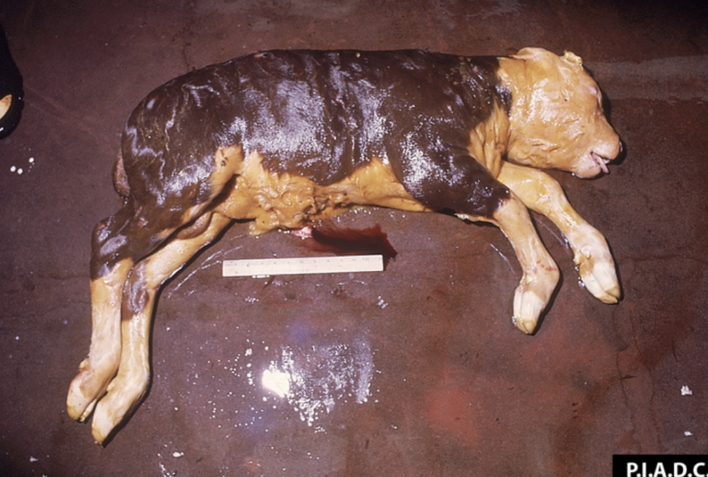
Abortion may be the only sign of leptospirosis in sheep and goats. Pigs and horses may also experience abortion if infected by this disease.

Leptospirosis causes delivery of dead pigs or poor survival of new born pigs. Kidney damage as a result of infection may also cause failure to thrive in pigs.

In horses, the eye is most commonly affected. The eyelids may be reddened, the horse will be sensitive to light and may blink frequently or clouding of the eye 

Leptospirosis Prevention 

Do not allow animals to be in contact with contaminated water or soil environments or infected wildlife, especially rodents. 

Leptospirosis vaccines are available for pigs, cattle and dogs. These vaccines help to prevent disease severity but may not completely prevent infection.

Always wear gloves, coveralls and gumboots especially if exposed to leptospira contaminated soil, water or infected livestock/other animals (during outbreaks)

Cover any cuts/wounds when handling animals. Avoid direct contact with urine, tissues and organs from potentially infected animals.

Avoid touching eyes, nose and the mouth after handling animals; and do so after proper hand washing. 

Practice good hygiene i.e. hand washing with soap and water after handling animals, pets or surfaces that might have pets’ excrement on it.

Keep livestock effluent (faeces and urine) separately, fence or keep stock away from effluent ponds, pits and waterways.

Routine vaccinations for animals whose vaccines are available i.e. dogs and cattle. ST – Ensure completion of treatment regimen for animals undergoing treatment.

Practice good rodent control practices by ensuring that crop and feed stores are rodent free e.g. by making sure no excess feed is lying around. 

Leptospirosis Warning 

Many animals carry leptospira bacteria but show no clinical signs. It is critical to strictly observe proper hygiene practices especially if an animal is sick e.g. aborted  

Lepospirosis is a notifiable disease as it can cause outbreaks. In case of suspicion, report to the nearest health or veterinary department immediately.

If you suspect leptospirosis: (1) In animals – contact your veterinarian immediately; (2) In humans – contact your physician immediately

Content provided by Zoonotic and Emerging Diseases Group (ZED) University of Liverpool , International Livestock Research Institute (ILRI), Zoonoses in Livestock in Kenya (ZooLink)

Foot and Mouth Disease

Inside of cows mouth suffering from foot and mouth disease.
Photo Credit ZooLink/ILRI

About F&M

Foot & Mouth disease is highly contagious and transmitted via inhalation or ingestion of virus from contaminated feeds, animals, meat, milk, semen and ticks. 

It can also be contacted by contamination from cars and people from infected areas. 

Symptoms

This disease causes severe lameness, blisters between toes, on teats and on tongue, high fever rapid loss of condition, long ropey strings of saliva. Abortion is common

Prevention

From age 2 weeks vaccinate every 6 months for Foot and Mouth disease. Different strains exist. Consult your veterinarian on the choice of vaccine.

To help animals heal, shade them from sun, give water, soft green grass and molasses for energy. Call Vet to prevent infection of blisters with antibiotics if necessary.

Disinfecting premises and motor vehicle tyres where F&M has occurred with suitable disinfectant.

Burn the feed and beddings of infected animals. Make sure to have foot dips with suitable disinfection for visitors. Make sure they use them!

Separate all sick animals and give them tender loving care.

NOTICE!

Foot and Mouth Disease is a NOTIFIABLE disease. This means you MUST report this disease to your DVO immediately to stop spread in community.

Kiswahili-Foot and Mouth Disease

About F&M

Ugonjwa ambukizi huenezwa kupitia kuvuta pumzi au kumeza virusi kutoka kwa lishe,wanyama,nyama, shahawa, kupe na mapagari kutoka maeneo yalioambukizwa.

Ugonjwa wa Foot and Mouth pia unaweza kuambukizwa kupitia magari yaliyo na uchafu wa maambukizi na watu kutoka maeneo ambayo yameadhiriwa.

Symptoms

Ulemavu mkali,uvimbe katikati ya vidole,kwa matiti na ulimi,joto kali, afya kudhoofika, kutokwa na mate ndefu na uavyaji mimba huwa kawaida. Mjulishe DVO.

Prevention

Kuanzia umri wa wiki ya pili mpe chanjo kila miezi 6 kwa ugonjwa ya Foot and Mouth. Pata ushauri kutoka kwa vet kuhusu chajo inayofaa.

Wasaidie mifugo kupona,waweke kwa kivuli wape maji,nyasi laini na molasses iwape nguvu.Dunga sindano ya intramuscular antibiotic izuie maambukizi uvimbeni

Eua na dawa inayofa majengo na matairi za gari ambazo magonjwa ya Foot and Mouth yametokea. Choma lishe na malazi ya mifugo walioambukizwa.

Choma lishe na malazi ya mifugo walioambukizwa. Hakikisha kuwa una dawa ya kueua miguu na dawa inayofaa wageni kutumia kujieua.Hakikisha wameitumia

NOTICE!

Foot and Mouth ni ugonjwa ambao unafaa kuripotiwa.Yamaanisha kuwa LAZIMA uripoti ugonjwa huu kwa DVO haraka iwezekanavyo ili kuizua kuenea katika eneo lenu

Content provided by Zoonotic and Emerging Diseases Group (ZED) University of Liverpool, International Livestock Research Institute (ILRI), Zoonoses in Livestock in Kenya (ZooLink)

Echinococcosis

Photo Credit ILRI/ZooLink Echinococcosis

Echinococcosis Description

Echinococcosis (hydatid disease) is a parasitic disease that affects animals and humans caused by several species of tiny tapeworms called Echinococcus

The tapeworms causing Echinococcosis are found worldwide. 

Echinococcosis Animal Symptoms

Echinococcosis can affect dogs, cats, sheep, cattle, pigs, and horses

Dogs and cats are termed definitive hosts because they are NECESSARY for the survival of the parasite while the other animals (livestock) are intermediate hosts

When dogs and cats are infected they do not show any apparent signs of illness 

In livestock the signs may range from no-signs to non-specific (nausea, vomiting, intermittent diarrhea, and weight loss)

In severe livestock infestations the parasites can cause fluid or masses in the abdomen, enlargement of the liver and abdomen, and difficulty with breathing.

In livestock, reproduction and production loses occur e.g. reduced growth and milk production, reduced birth rate and losses due to condemnation of organs at PM

Severe illness in animals is exhibited by multiple cysts being found in the brain, kidneys, bones, or testes 

Without control measures, infection rates can be very high in livestock, cats and dogs, with associated significant incidence in humans.

Echinococcus Animal transmission

Dogs and cats are infected by eating infected meat (meat with Echinococcus cysts) from livestock (sheep, goats, pigs and cattle)

Livestock (sheep, goats, pigs and cattle), are infected after ingesting Echinococcus eggs passed in the feces of dogs and cats

The Echinococcus eggs can survive for several months in the environment (e.g., pastures, gardens) especially in warm, moist conditions.

EchinococcusHuman Symptoms

Clinical signs in humans depend on where in the body the cyst develops, the size and number of cysts e.g. the larger the cyst the severe the symptoms

When the Echinococcus cysts are in the liver, symptoms experienced include: Abdominal pain, nausea and vomiting 

When the Echinococcus cysts are in the lung (s) clinical signs include: chronic cough, chest pain and shortness of breath

Other signs depend on the location of the hydatid cysts and the pressure exerted on the surrounding tissues. Non-specific signs include anorexia, weight loss and weakness.

The severe form of the Echinococcosis occurs when multiple cysts of Echinococcus occur in the brain, kidneys, bone, or testes causing more severe illness

Echinococcus Human transmission

Humans are infected through eating contaminated food, water or soil, or through direct contact with infected faeces of livestock, dogs and cats.

Echinococcus Prevention

Preventing access of dogs to livestock carcasses or slaughter wastes from farms, households, abattoirs or butchers can help control the spread of Echinococossis

If cysts are detected at meat inspection, infected farms or communities should be targeted and control measures put in place.

Vaccinating sheep (or other livestock) to protect against the development of the larval stage of Echinococcus

Wash all fruits or vegetables thoroughly before eating them, particularly those picked in the wild or directly off the ground. 

Always wash your hands with warm water and soap after handling dogs, cats and livestock

Echinococcus Action

Regularly examine and treat dogs and cats that are routinely exposed to livestock (e.g. sheep, goats, pigs and cattle) 

If you suspect echinococcosis: (1) In animals – contact your veterinarian immediately; (2) In humans – contact your physician immediately

Content provided by Zoonotic and Emerging Diseases Group (ZED) University of Liverpool, International Livestock Research Institute (ILRI), Zoonoses in Livestock in Kenya (ZooLink)

Cysticercosis

Photo Credits ILRI/ZooLink Cysticercosis Tongue Cysts

Cysticercosis Description

Cysticercosis is a parasitic infection caused by pork tapeworm cysts in different human body parts e.g. muscles, skin, eyes, brain

Taeniasis is infection with an adult pork tapeworm in the human intestine; this can result in abdominal pain and weight loss

Cysticercosis is found worldwide. Infection is highest in many parts of the developing countries due to poor sanitation and close proximity to infected pigs

Cysticercosis Transmission

In the human intestines the young pork tapeworm come out of their cysts and develop into adult tapeworms which then lay eggs in the human intestines

Eating food and water contaminated by human stool can transmit eggs to pigs. In the pig eggs develop into larvae and form cysts in muscles and tissues

Cysticercosis Animal Symptoms

Symptoms of cysticercosis are uncommon in pigs but occasionally you will see hypersensitivity of the snout, tongue paralysis, seizures, fever and muscle stiffness 

At slaughter the cysticercosis cysts are mainly found in the skeletal or heart muscles, liver, heart and brain of pigs.

Cysticercosis Human transmission

Human tapeworm infection (taeniasis) occurs after ingesting raw or undercooked pork

Humans may carry the pork tapeworm in their intestine (taeniasis), often without symptoms

The infected person will shed the pork tapeworm eggs in stool. Cysticercosis will develop in whoever consumes the contaminated fecal matter. 

People get cysticercosis through eating food (mainly vegetables), water, or surfaces contaminated with stool or through self-infection due to poor hygiene.  

People living in the same household with someone who has a tapeworm infection have a much higher risk of getting cysticercosis than people who don’t.

Cysticercosis Human Symptoms

Taeniasis is usually characterized by mild and non-specific symptoms e.g. abdominal pain, nausea, diarrhoea or constipation within 6–8 weeks after infection  

In cysticercosis, people may remain asymptomatic for years i.e. symptoms occur months to years after infection, usually when the cysts start dying.

Cysts in the muscles generally do not cause symptoms. However, you may be able to feel lumps under your skin. The lumps sometimes become tender. 

Although rare, cysts may float in the eye and cause blurry or disturbed vision. Infection in the eyes may cause swelling or detachment of the retina. 

Neurocysticercosis (cysts in the brain, spinal cord) mainly cause seizures, chronic headaches, seizures, dementia, changes in walking. This can result in death.

Cysticercosis Prevention

Avoid unclean foods, don’t eat uncooked foods while traveling or visiting friends and relatives, and always wash and peel all raw fruits and vegetables before eating

Educate family members, children and friends about proper food handling, good personal hygiene, and proper hand and body sanitation.

Avoid eating raw or undercooked pork to avoid intestinal infection (taeniasis)

Wash your hands with soap and warm water after using the toilet, changing diapers, and before handling food and properly handle and prepare foods 

Use good food and water safety practices while visiting friends and relatives. Ensure that to drink only boiled or bottled or treated water. 

Preventing or treating taeniasis in people and preventing animal exposure to human stool can reduce cysticercosis in livestock 

Avoid free range pig farming and practice confining pigs to pig-pens to avoid their exposure to human stool which is a source of their infection/re-infection.

Cysticercosis Action

If you suspect that you have any symptoms of cysticercosis or taeniasis, contact your health care provider immediately

If you suspect cysticercosis: (1) In animals – contact your veterinarian immediately; (2) In humans – contact your physician immediately

Content provided by Zoonotic and Emerging Diseases Group (ZED) University of Liverpool, International Livestock Research Institute (ILRI), Zoonoses in Livestock in Kenya (ZooLink)