The Water Cycle

Credits: Biovision-Infonet

Every living creature and vegetation must have water in order to survive.

Only 3% of all water on Earth is fresh water and 68% of the water is frozen ice on the North and South Poles.

All humans and most animals as well as nearly all vegetation can only survive on fresh water free of salt and minerals.

Water for irrigation must also be fresh water and applied sparingly by flood, furrow or drip irrigation, otherwise the irrigated soil will turn saline and unproductive for many years to come.

The other 97% of the water on planet Earth is saline seawater in which whales, fish, corals and plankton flourish. Slightly salty water may be used for watering livestock and other animals, although fresh water is healthier.

Nearly half of all deep boreholes are dry or contain saline (brackish) water with minerals harmful to humans, animals and plants.

Where fresh water is pumped up from very deep boreholes it is called mining fossil water because the water withdrawn cannot be replaced by rains.

Therefore, there are only two sustainable solutions to the world’s increasing demand for water, namely rainwater harvesting and water conservation.

Fresh water sources are replenished in a water cycle through the following activities: 

  1. Evaporation is an almost invisible vapour rising from water surfaces.
  2. Transpiration is evaporation from vegetation and soil surfaces. 
  3. Precipitation from clouds falls as fogs, mists, rains, hails and snow.
  4. Rainwater run-off is rainwater running off all kind of surfaces.
  5. Drainage is the ability to drain excess water away from catchments.
  6. Infiltration is the movement of water into the soil from the surface. 
  7. Percolation is the movement of water through the soil to the underground layers.
  8. Permeability is the rate at which water penetrates through soils down into the underground water table. 
  9. In situ storage is storage of water in the voids between particles of soil and sand.
  10.  Subsurface flow is a flow of water in the voids of soil and sand particles.  

Water can be harvested at several stages in the water cycle, such as: 

  1.  Fog screens placed on hills, mountains and near the sea for domestic water. 
  2.  Gutters attached to roofs for domestic water. 
  3. Garlands of stone gutters on rock outcrops for domestic water. 
  4.  Soil bunds and trenches on farmland for crops and animals. 
  5. Diversion trenches from roads, rocks and hillsides for seasonal flood irrigation.
  6. Hand dug wells in shallow ground water to supply water for all uses. 
  7.  Subsurface dams, weirs and sand dams to increase the yield of hand dug wells situated along seasonal water courses. 
  8.  Boreholes drilled into deep ground water where none of the above options are replicable, although expensive and only 40% may not supply fresh water.  

The quality of water from all these sources can be tested by either a portable testing kit or by a laboratory if the samples can be delivered before deteriorating due to heat over long distances.

Contaminated water may be treated by several methods, such as SODIS (Solar disinfection), boiling, water filters, crushed seeds from the Moringa tree, ultra-violet rays (UV) or artificial chemicals.

Rainwater Harvesting

Credits: Biovision-Infonet

No matter how uneven or how long a roof is, gutters can be hung with the required gradient of 1 cm drop for every 100 cm length (1:100), when a splash-guard is nailed onto the roof
(c) E. Nissen-Petersen, Kenya
Gutters fitted into hangers tied to a splash-guard nailed onto an uneven roof
(c) E. Nissen-Petersen, Kenya
A garland of gutters at Kasigau
(c) E. Nissen-Petersen, Kenya

Introduction

Rainwater running off land surfaces can be harvested, stored and utilized using a technique called rainwater harvesting instead of being wasted in rivers, lakes and the sea.  

Rainwater harvesting consists of 5 components: 

  1.  Rainfall 
  2.  Catchment areas, also called watersheds, onto which the rainwater falls.
  3.  Gutters, or conveying channels, to bring rainwater from a catchment area to storage reservoir. 
  4.   Storage reservoirs can be tanks, ponds, dams and in situ storage in sand and soil.
  5.  Retrieval water is extracted from reservoirs either by gravity or by pumps and lifts.

A rural homestead should preferably have the following variety of structures for harvesting rainwater to avoid water shortages during dry seasons: 

1) A roof catchment system for clean domestic water that consists of gutters fixed to the roof which drain the rainwater into a storage tank. The size of a storage tank depends mainly on the financial capacity of the owner and to a lesser degree on the size of the roof and the volume of rainfall. However, the ability to supply sufficient water during years with drought depends on the size of the roof and the tank.

2) A pond or an earth dam for watering livestock and garden irrigation can be excavated by hand or animal drawn implements at a low place in the farmland where rainwater flows, or accumulates, during rainy seasons. Ponds and dams can initially be built small and enlarged during the following dry seasons until they might supply water throughout the years. 

3) A hand dug well may supply water if sunk into shallow ground water, e.g. downstream of an earth dam or near evergreen trees growing on riverbanks. 

In situ harvesting of rainwater in farmland increases the yield of crops and will often determine whether there will be anything to harvest at all. Most farmers know and apply some of the techniques of soil conservation that make rainwater percolate into the soil instead of eroding the farmland. Among several techniques, the following are being used by many farmers:

  1. Contour planting in horizontal ploughing lines.
  2. Contour trash lines with grass and farm waste.
  3. Contour ridges that develop into terraces. 
  4. Contour bunds of stones that develop into terraces.
  5. Bench terraces that develop slowly from contour ridges.
  6. Micro catchments with U and V shaped soil bunds for growing grass and crops and trees.
  7. Trapezoidal bunds with a farming area of 1,350 m2 (1/3 acre) for grass and crops.
  8. Cut-off drains to discharge surplus rainwater run-off into gullies with check dams and streams.
  9.  Check dams of stones and vegetation to heal eroded gullies.
  10.  Check dams of stones and vegetation in valleys.

Pig Information Source

Credits:Biovision-Infonet

  • AIC Documentation Unit, Kenya
  • Feeding organic pigs, A handbook of raw materials and recommendations of feeding practice, 2002. University of Newcastle
  • Livestock production extension manual (MoLD – Kenya) (2008).
  • Wabacha, J.K., Mribei, J.M., Mulei, C.M., Kyule, M.N., Zessin, Z.H. and Oluoch-Kosura, W. (2004). Characterization of smallholder pig production in Kikuyu Division, Central Kenya. In: Preventive Veterinary Medicine 63 (3) pg 183-195.
Last updated on:

Wed, 10/02/2019 – 08:49

Pigs Reproductive Disorders and Diseases

Credits:Biovision-Infonet

These include, Brucellosis, mastitis, leptospirosis, endometritis and agalactia. 

Anaphrodisias 

When the sow does not come on heat 

Symptoms: 

  • No heat signs
    This could be caused by low body weight due to poor feeding, overweight, mineral deficiency, intestinal worms, chronic disease, the animal has just given birth, heavy infestation with parasites

Prevention:

  • Improve feeding of mineral-rich feeds. 
  • Regularly deworm your animals. 
  • Allow the female to stay with the boar.

Treatment:

  • Gilts should not be treated at all because they may show anaphrodisias after every litter
  • Sows should be treated on the same day as weaning, and in very severe cases, 3 weeks after weaning
  • Repeating treatment is of no use

Parvo (smedi) (virus)

Symptoms:

  • Birth of a small litter at the normal time due to Early Embryoric Death (EED) with mummies of different ages together with fresh or macerated dead foetuses and weak living piglets which die in few days.

Prevention and treatment

  • No therapy 
  • Vaccination of all gilts and sows before pregnancy

Leptospirosis (Bacteria)

Symptoms:

  • Fever, anorexia, diarrhoea, bloody urine, nervous symptons caused by meningitis.
  • Abortion in last three months of pregnancy. 
  • In sows which are affected later, weak piglets are born. 
  • Mummified and macerated foetuses are common in the litters. 
  • Infertility associated with venereal spread may be responsible for repeat breeders.

Prevention and treatment:

  • Elimination of mice and rats and other rodents. 
  • Vaccination and hygienic measures. Vaccines are not available for all types of the diseases and vaccination may not prevent bloody urine. 
  • Treat all sows with injection or streptomycin before serving. 
  • Use antibiotibics especially streptomycin for all ages.


Brucellosis

Symptoms:

  • Anorexia, fever, stiff legs, occasional lameness, early abortion (returns to oestrus 5 – 8 weeks after service as a result of infection of service)
  • Infection later in pregnancy gives rise to litter with mummified, still born or weak piglets. 
  • Bloody vulva discharge and endometritis. 
  • Retained placenta. 
  • Boars usually develop orchitis (inflammation of one or both testicles) and epididymitis within seven days of infection. 
  • The testicles are swollen and painful and permanent sterility can be the result

Prevention and treatment

  • Prevention is based on hygienic measures and purchase of stock from clean herds only. 
  • Never treat by antibiotics. 
  • No treatment/vaccination is 100% effective
  • Slaughter all animals and do restocking. Restocking should be after one month.

Uterine Prolapse

Symptoms:

  • The appearance of the uterus outside the vulva.

Prevention and treatment:

  • Uterine prolapse reduction is often not possible since it is very traumatic and the best therapy is to amputate the whole uterus. However 50% of sows do not survive this operation, therefore slaughter should be considered.

Mastitis

Bacteria infection causes an inflammation of the mammary organ and results in changes in milk production. These bacteria enter the wounds in the udder. 

Symptoms

  • Swollen, hot and painful udder. 
  • Absence or reduction of milk in the affected udder. 
  • Sow refuses to suckle her piglets. As a result, piglets squeal due to hunger. 
  • Sow has depression and often fever.

Prevention:

  • Provide adequate bedding 
  • Keep pig pens clean, dry and free of sharp objects, clip milk teeth of baby pigs. 

Treatment:

  • Gently massage the affected udder with lukewarm water. 
  • Do not allow the young to suck milk from the infected sow. 
  • Remove the milk from the infected udder and discard. 
  • Separate sow from piglets and reduce access to teats (allow a few piglets to suckle at a time). If possible, foster piglets to lactating mothers. 
  • Use antibiotics. Inject penicillin-streptomycin into the muscle of hip or neck. 

Endometritis (bacteria)

Symptoms

  • Vulval discharge of vaginal or uterine origin during urination. 
  • In case of metrititis, fever and agalactia may occur.

Prevention and treatment

  • Antimicrobial by injection or locally by irrigation of the uterus and vagina, oxytocin can be given to stimulate uteric contractions once or twice a day. 
  • Hygiene.

Diseases and Disorders of Digestive TractScouring

  • Hygienic measures should be taken to avoid or minimize scouring incidence.
  • Regular deworming should also be done as a control measure to scouring
  • Feed changes should be gradual and not drastic to avoid scouring

Diarrhoea/Enteritis1) Birth diarrhoea

Symptoms

  • Acute diarrhoea in piglets is watery, yellowish grey and within a very short time piglets become thin with sunken eyes due to dehydration. 
  • Death can occur within 2 – 3 days. Death in piglets can occur even within one day before any sign of diarrhoea is observed.

Prevention 

  • Keep pens, feed and watering troughs clean.
  • Separate affected animals from healthy animals.
  • Do not change abruptly an animal’s ration. 
  • Make sure that piglets have sufficient colostrum within 36 hours of birth

Treatment

  • Vaccination
  • Plenty of drinking water with electrolytes
  • Use of antibiotics.
  • Herbal medicine: Fresh leaves of guava or star apple (Sapodilla) This will treat the symptoms only. 

2) Red diarrhoea or Clostridial enteritis

Symptoms:

  • Acute haemorrhagic or necrotic inflammation of the gut during the first and second week of birth. 
  • In acute cases the diarrhoea is watery with an orange-red colour due to blood and is often bubbly. 
  • In later stages, shreds of dead tissues can be found. 
  • Piglets are weak and don’t suckle. They get pale and die within a few days.

Prevention and treatment

  • Vaccination
  • Make sure that piglets have sufficient colostrum within 24 hours of birth 
  • Keep the pigs warm
  • Cleanliness
  • Contact the vet

3) Transmissible Gastroenteritis

Transmissible gastroenteritis is a common viral disease of the small intestine that causes vomiting and profuse diarrhoea in pigs of all ages. It spreads rapidly. Piglets less than one-week old rarely survive the disease. Symptoms:

  • Very high mortality mainly in piglets upto 14 days old. 
  • The piglets often vomit and have severe greenish-yellow watery diarrhoea, dehydration. 
  • In sows the diarrhoea is greyish, they vomit, and abortion may occur. 
  • In fatteners the symptoms are like those in sows.

Prevention and treatment:

  • Give electrolytes to piglets and keep them warm. 
  • Antibiotics prevent secondary infection but don’t provide a cure.

4) Fat diarrhoea

Symptoms:

  • Faeces are pasty and fatty, white or yellowish

Prevention and treatment

  • Remove creep feed for a few days. 
  • Use antibiotics as prescribed by a veterinary doctor.

5) Post diarrhoea

Symptoms:

  • Diarrhoea with no traces of blood. 
  • Death from dehydration or blood poisoning (septicaemia).

Prevention and treatment:

  • Increase creep feed before weaning. 
  • Avoid stress to piglets by not mixing piglets from different litters. 
  • Good hygienic measures include roughage in diet.
  • Use antibiotics as prescribed by a veterinary doctor.

Salmonellosis

Symptoms:

  • High fever, dullness, anorexia, weakness, nervous symptoms.
  • Bluish-red colouring of the ears, limbs and the centre of the belly. 
  • Bloody spots all over the body. 
  • Wasting and persistent greyish diarrhoea sometimes mixed with blood and shreds or necrotic material from the gut.

Prevention and treatment: 

  • Normal hygienic measures, pelleted feed, thorough cooking of the swill. 
  • Remove feed for two days and provide clean water. 
  • Use antibiotics as prescribed by a veterinary doctor.

Swine Dysentry

Known by a number of names, including bloody diarrhoea, hemorrhagic enteritis bloody scours and black scours. It affects pigs of all ages, sometimes causing death.

Symptoms:

  • In acute cases wasting and passing of diarrhoea containing varying amounts of mucus, blood and necrotic material. 
  • Fever. 
  • In chronic cases, pigs have greyish or brownish faeces, rough hair coat and low growth rate.

Prevention 

  • Delay reusing the pens of infected animals.
  • Disinfect pens. 

Treatment: 

  • Some herbal medicines (Moringa tree leaves) can be used to relieve the symptoms of diarrhoea and dehydration 
  • Use antibiotics as prescribed by a veterinary doctor

Post weaning syndrome (oedema disease)

Symptoms:

  • Development of oedema and nervous signs. 
  • Paleness peculiar squeaky voice. 
  • General incoordination and loss of balance. 
  • Sudden death.

Prevention and treatment

  • Affected piglets should be weaned by removal of sow and placed on low-level diet. 
  • Avoid stress. 
  • Hygiene. 
  • Include roughages in diet. 
  • Use antibiotics as prescribed by a veterinary doctor

Diseases of the Respiratory Tract

These include, influenza, pseudorabies, pneumonia, atrophic rhinitis and pasteurellosis.

 Influenza

Symptoms:

  • Affected animals are apathetic. 
  • Anorexia
  • High fever, coughing and sneezing, difficulty in breathing
  • Red eyes with discharge
  • Loss of condition.

Prevention and treatment:

  • Good ventilation. 
  • Vaccination. 
  • No specific treatment. To prevent secondary infection, use antibiotics as prescribed by a veterinary doctor .

Pleural Pneumonia

Symptoms:

  • Acute cases show anorexia, high fever, laboured respiration, red or blue colouring of ear-tips, belly, legs and end of tail. 
  • Death within 4 – 6 hours of onset of clinical symptoms. 
  • Blood stained froth from mouth or nose. 
  • Abortion. 
  • In chronic cases, anorexia, coughing and depressed growth rate.

Prevention and treatment:

  • Vaccines only prevent mortality. 
  • Use antibiotics as prescribed by a veterinary doctor.

Atrophic rhinitis (inflammation of the nose)

Symptoms:

  • Sneezing in younger pigs. 
  • Shortening or deviation of upper jaw – poor growth

Prevention and treatment:

  • Hygiene 
  • Use antibiotics as prescribed by a veterinary doctor

Pasteurellosis

Symptoms:

  • Coughing
  • Breathing through the mouth.

Prevention and treatment:

  • Hygiene.
  • Use antibiotics as prescribed by a veterinary doctor 

Porcine Respiratory and Reproductive Syndrome (PRRS)

Symptoms:

  • Blue ears and forced breathing
  • Lactating sows have rough hair coat. 
  • Gilts have problem getting on heat and early abortion. 
  • High mortality in weaners 
  • In boars, high percentage of defective sperm and less libido 

Prevention and treatment: 

  • Use of antibiotics as prescribed by a veterinary doctor. 
  • Vaccination helps but it is so expensive that it may not be cost effective.

Disease Causing Problems in WalkingArthritis

Symptoms:

  • Inflammation of joints. Thick soft joints.

Prevention and treatment: 

  • Use antibiotics as prescribed by a veterinary doctor.

Streptococcal infections
 

Symptoms:

  • Septicaemia (blood poisoning) which may cause immediate death.
  • Young pigs rarely recover
  • Sudden death in older pigs.
  • Fever, nervous signs and arthritis mostly in weaners and fatteners.

Prevention and treatment: 

  • Proper hygiene. Wash sow before it enters the farrowing pen. 
  • Use antibiotics as prescribed by a veterinary doctor.

Greasy Pig Disease

Symptoms:

  • Thin, pale brown flakes on the skin surface. 
  • Wet skin covered with crusts. 
  • Rough wet and reddish skin below the crusts, no itching
  • Death may occur

Prevention and treatment:

  • Avoid fighting among pigs by mixing pigs from different pens. 
  • Teeth clipping, soft bedding 
  • Hygiene. Wash sows before entering farrowing pen. 
  • Use antibiotics as prescribed by a veterinary doctor.

Nutritional Disorders

Anaemia

Symptoms:

  • Pale skin, weak piglets with high respiratory rate. 
  • Jaundice
  • Blood stained faeces.
  • Early death

Control and treatment:

  • Provide iron injection or oral iron- paste containing iron, depending on the situation – consult a vet or an advisor
  • Feeding compost- must be of good quality and supplied daily. Compost of poor quality may contain bacteria.
  • Wood ash can also be put into the pen. This will not provide iron, but it does contain other important minerals.

Other Diseases

Foot and mouth disease (FMD)

Foot-and-mouth disease is an acute, highly contagious, viral disease of animals with hooves, such as cattle, water buffalo, goats and pigs. 

Symptoms:

  • Sudden onset of severe lameness, fever, formation of vesicles on coronary bands. 
  • Blisters can be found on thin-skinned areas like udder, teats, anal area and eyelids. These blisters rupture within one day. 
  • There may be frothy saliva, anorexia, sometimes hooves become loose and fall off.
  • Sows may abort.

Prevention and treatment:

  • Vaccination
  • Quarantine 
  • Proper cooking of swill.
  • Slaughter and burial. 

African Swine Fever (viral)

Symptoms:

  • Lesions on the body 
  • Fall in temperature before onset of clinical signs. 
  • Reddening of skin and ears. 
  • Incordination of hind limbs. 
  • Constipation diarrhoea, anorexia. 
  • The animal dies the next day after the attack. 95 – 100% mortality.
A pig with African Swine Fever, showing a bluish-purple colour and bleeding under the skin.
(c) S. Gikonyo, Kenya

Prevention and treatment:

  • Quarantine. 
  • Boiling of swill. 
  • Restriction of movement of meat from infected areas
  • Vaccination
  • Disinfection
  • No therapy (treatment)

Anthrax (bacterial)

Rare in pigs and associated with contaminated feed containing meat.
Symptoms: 

  • Oedema and swelling of the neck region. 
  • Dysponea (breathing difficulty). 
  • Fever, anorexia and passage of bloody faeces. 
  • Sudden death.

Prevention and treatment:

  • Use antibiotics (penicillin) as prescribed by a veterinary doctor.
  • Thorough disinfection of the farm and burning of carcasses.

Potato Seed Production Info Source Links

Credits: Biovision-Infonet

  • Anon. (2006). Select the best: Field guide for positive seed potato selection. CIP Sub-Saharan Office, Nairobi
  • Gildemacher, P., P. Demo, P. Kinyae, M. Wakahiu, M. Nyongesa and T. Zscocke (2009). Select the best. Positive selection to improve farm saved seed potatoes. Farm field aid. 2nd. Edition. CIP & CTA. ISBN: 978-92-9060-362-7
  • Kabira, J.N, M. Macharia, M.W. Karanja and L.M. Muriithi (2006). Potato Seed: How to grow and market healthy planting material. KARI Technical Note No. 20, April, 2006, Kenya Agricultural Research Institute
  • Kinyua, Z.M and J.J. Smith (2007). Multiplication of disease-free seed potato in seed plots. KARI Advisory Bulletin, March 2007, Kenya Agricultural Research Institute
  • Nyongesa, M., C. Lung’aho, C. Kinyae, P. Wakahiu, M. Karinga and J. Kabira (2008). Production of food (ware) potatoes. KARI information brochure series/35/2008, Kenya Agricultural Research Institute
  • Nyongesa, M., C. Lung’aho, P. Wakahiu, J.N. Kabira, P. Gildemacher and G. Forbes (2008). Control of late blight on varieties Asante and Tigoni. Kenya Agricultural Research Institute/International Potato Centre, SSA, Nairobi

Potato Seed Production

Credits: Biovision-Infonet

Potato field
(c) A.A. Seif
Healthy Mavuno potato
© A.A. Seif

Introduction

Second only to maize, “Irish” potatoes play a very big role in the Kenya food security situation. Potatoes can be grown within a growing period of 3 months, produce good yields of highly nutritious food and be stored for some months under the right conditions.

However growing potatoes in mixed cropping systems as currently practised by many small scale farmers where volunteers from previous crops are not weeded out has by now managed to spread serious seed borne plant diseases to most potato growing areas.

This has resulted in declining yields and again undermined the possibility of good food security in These areas. 

In order to clean up the disease situation and to increase clean side production it is necessary to understand exactly what needs to be done (for more articles on clean seed potato production please also see TOF June 2010 Farmers can also grow their own seed and share with neighbours, if good field hygiene is observed as described in this article. 

On-farm production of potato seed tubers by smallholder farmers in small seed-plots

Principle of seed plot system

This involves maximising tuber production per unit area of limited, disease-free land through high-density planting in a seed plot and also separation of seed and ware (larger tubers meant for consumption) potato production.

The system uses 50% less land than the ware production to meet on-farm seed tuber requirements 

Land requirement and site selection

Commercial Opportunity: 

In Kenya there is a very large commercial opportunity in the production of certified seed potatoes.

The demand for and commercial price of clean potato seed tubers is far higher than potatoes for the food market. Mid 2010 it was established that Kenya only produces about 1% of the potato seeds needed every year by potato Farmers countrywide.

Clean seed is the best start for a good potato crop yield as most potato farmers know or have discovered. 

So if you have an extra piece of land free of bacterial wilt and other potato plant diseases, it might be a very good ideal to pay a visit to KEPHIS (Kenya Plant Health Inspectorate Service ) and find out from them how to go about such a venture.

The production of certified seeds can easly be carried out in farmers fields, but will need inspection and certification by KEPHIS officers to be able to attract the nice high price tag that currently is offered for such clean seed.

Choose your plot for potato seed production with care:

  • It should not have had any Irish potatoes, capsicums, eggplants (brinjals) and tomatoes grown in it for the last 5 years.
  • Preferably it should have had a dense mat of grass growing on it for at least one year during the last 5 years.
  • Other probable sites for seed plots include virgin land and land that has been under fallow for 2-3 seasons.
  • It should not have been given agricultural lime for the last 5 years
  • The plot should be located in full sunshine in highland areas, and in half shade (for example shaded half the day) in hotter areas.
  • The seed plot should not be situated in low-lying or water-logged areas where run-off water flows into it.
  • There should be a good supply of well composted farm yard manure applied just before planting the certified seed potato tubers. If the amount of manure is not very much, it should be applied in the trenches dug for planting tubers.
  • If Lantana camara bushes grow in your neighbourhood it is very beneficial to also mix in leaves of lantana in the potato planting rows to repel insects.

Sources of seed tubers

Clean seed tubers can be obtained from KALRO centres at Tigoni, Embu, Kitale and Oljoro Orok, identified seed growers who work with Ministry of Agriculture, and from authorised farmers groups who produce quality seed with advisory inspection support from Kenya Plant Health Inspectorate Service (KEPHIS).

However, commercial clean seed is expensive and mostly not available. Therefore, for most farmers, positive seed selection and seed plot system for seed production offer an immediate low-cost alternative, provided it is done correctly

Positive seed selection technique

This is a procedure where any diseased plant particularly infected with bacterial wilt or viruses is removed from the field immediately it is noticed and the remaining healthy-looking plants at flowering are marked with sticks or pegs. The marked or selected healthy-looking plants should be checked weekly and those which have developed disease symptoms should be un pegged and removed from the field. Pegged plants should not be near (at least 1 metre away) where diseased plants were removed. 

  • These pegged plants are considered to be positively selected seed potatoes. The removed diseased plants with bacterial wilt including their tubers should be put in a 1-metre deep pit and covered with clean soil or be burnt. Also wilt infected plants and tubers should not be put in compost heap.
  •  Harvest the pegged plants separately and first before the rest of the field to get seed tubers for the next planting. This procedure is in contrast with the usual farmer practice where all plants in the field are harvested together and seed is picked from the harvest for planting in the following season

Guide to knowing healthy and diseased potato plants

How a healthy plant looks:

A healthy plant is vigorous, has many stems, dark-green leaves and at harvest, has more seed-sized of 35-55 mm than smaller ones below 25 mm in diameter. 

Healthy Mavuno potato
(c) A.A. Seif
Potato variety ‘Asante’
(c) A.A. Seif
Potato variety ‘Tigoni’
(c) A.A. Seif
Potato crop flowering
(c) A.A. Seif

Sudden wilting of plants even when it is wet. Entire plants can wilt quickly without yellowing.

There is a dark brown colour in the inner section of the stem. A white creamy liquid oozes out when the stem is cut near the base.

Tubers are black or have black eye buds. Heavily infected tubers have soil stuck to the tuber eyes and on cutting of the tubers rotting from the vascular ring is observed

Bacterial wilt (rotting of vascular ring)
(c) A.A. Seif
Bacterial wilt symptoms on potato plants. The first visible symptom on foliage is a wilting of the leaves at the ends of the branches during the heat of the day with recovery at night. As the disease develops, a streaky brown discolouration of the stem may be observed on stems 2.5 cm or more above the soil line, and the leaves develop a bronze tint.
(c) Mauritius Sugar Industry Research Institute. Reproduced from the Crop Protection Compendium, 2004 Edition. (c) CAB International, Wallingford, UK, 20

How a virus infected plant looks:

Plants are stunted (dwarfed). Leaves may have mosaic pattern (yellow and green). Leaf margins may be curled or rolled up and may be thick. In dry season, green aphids may be found on lower leaf surfaces. Tubers produced are small

Potato Leaf Roll Virus 1
(c) A. A. Seif

Seed plot system

Land preparation, planting and crop management

Mark out beds of 2.0 m width and a sufficient length depending on your seed needs. Plant in furrows at a spacing of 30 x 30 cm at planting depth of 15 cm.  A 90 m2 seed plot (2 x 45 m) can provide enough material to plant 0.4 ha (1 acre) of ware potatoes (4000 m2).

Hand weeding is recommended. Weeding should be done on appearance of weeds until there is sufficient potato growth to suppress weed growth.

 Seed plot should be weekly inspected to monitor crop performance and particularly occurrence of pests (aphids, potato tuber moth) and diseases (bacterial wilt, viruses, early and late blight).

Aphids can be easily controlled by spot insecticide application (consult area extension agricultural officers or the nearest KALRO centre for advice).

In case of potato tuber moth ridging is recommended. Any plant with bacterial wilt and/or virus infection should be uprooted and be destroyed and should not be used as animal feed as they may be brought back to the field in form of manure.

Put 2 hands-full of ashes or 1 hand-full of lime in the hole after removing a wilted plant.

Early and late blight can be effectively controlled by using appropriate fungicide immediately when noticed (consult area extension agricultural officers or the nearest KALRO centre for advice)

Potato mother plants widely spaced for production of basic seed
(c) A.A. Seif
Potato mother plants widely spaced (1.5×0.60m) for production of basic seed
(c) A.A. Seif

Harvesting, grading and storage

 On maturity, when leaves begin turning yellow, remove potato vines (haulms / stems) about 2 weeks before harvesting to harden tuber skin. Harvest when the soil is dry. Avoid injuring the tubers.

  • Seed tubers must be unbruised measuring 2.5-5.5 cm in diameter. Larger tubers should be sold off as ware potatoes.
  • Selected seed tubers should be stored in diffused light and ample aeration. Do not store seed tubers in dark stores used for ware potatoes.
  • Store tubers of each variety separately. Do not store seed tubers in gunny bags.
  • Spread the seed tubers thinly on the floor or in wooden crates.
  • Cover with branches of Lantana camara.
  • Farmers in Naivasha have discovered that storing seed potatoes under saw dust keeps away tuber moths and keep the potatoes seeds longer.

Usage of seed plot tubers

Tubers harvested from seed plots are divided into 2 lots: one lot is used to establish a new seed plot (for not more than 3 seasons) as described above and the other lot is used for production of ware potato in main fields. 

Seed plot protection

  • Fence the seed plot area to restrict movement that can cause contamination.
  • Plant maize around the seed plot as a border crop to minimize aphid infestation. Plant at least 4 rows of maize 2-3 weeks before seed potatoes.
  • Clean /and disinfect all tools, feet and shoes before entering the seed plot area.
  • Use a household bleach (sodium hypchlorite) (e.g. JIK(r)) at the rate of 50 ml in 1 litre of water.

Goose Pests

Credits: Biovision-Infonet

NEMATODES (worms)

The main problem with geese in EA is that they are susceptible to gizzard worms which kill them if left untreated. Gizzard worms are a very common parasite and geese should be wormed at least twice a year with 1ml Levamisole 7.5% (consult your pharmacist who will advise you of similar products available.

Never use more than the recommended dose as it is easy to overdose and this may prove fatal. This treatment is not suitable for goslings 10 weeks and younger.

Alternatively, use Flubenvet which is a multipurpose wormer and covers gizzard worm. It is a powder which sticks readily to feed. The correct dose (on the pack) should be administered for 7 days.

It is suitable for goslings and it is adviseable to treat a hen when she starts sitting, as well as the gander.
It has been said that nematodes, or roundworms as they are commonly called, constitute the most important group of helminth parasites of poultry.

With geese, Ascaridia are generally not a problem but various species of Capillaria and Heterakis can cause problems. The most common nematode in geese is Amidostomum anseris.

Symptoms

The usual symptom of worm infection in geese is lethargy. The presence of eggs or worms in either the faeces or in any organ, as revealed upon autopsy, will confirm a worm infection. Amidostomum anseris infects the horny lining of the gizzard and sometimes the proventriculus. It causes dark discoloration of the gizzard and a sloughing off of the lining.

Prevention/Treatment

The first principle in controlling nematode infections is to practice good management.

For geese on range, it is essential to rotate pastures 3-4 times a year and to change the pastures every year so as to break the cycle of nematode re-infection.

For geese in confinement, the litter should be changed regularly and the building washed and disinfected with insecticide after each flock of geese. It is important not to mix young and old geese together, nor to follow old geese with young geese who are much more susceptible to nematode infections.

A number of anthelmintic drugs are available to treat nematode infections. For the control of Amidostomum anseris in geese cambendazole, pyrantel, mebendazole and fenbendazole have each been shown to be effective. 

The following nematodes have been isolated from the small intestine of the goose: Echinura uncinata, Epomidiostomum uncinatum, Ascaridia galli, Capillaria anatis, Capillaria bursata, Capillaria annulata, Capillaria anseris, Capillaria caundinflata, Capillaria obsignata, Heterakis dispar, Heterakis gallinarum, Strongyloides avium and Trichostronglus tenuis. The nematode Syngamus trachea has been isolated from the respiratory tract of the goose.

TAPEWORMS

Over 1 400 species of cestodes or tapeworms have been noted in wild and domestic birds and for many an intermediate host has been identified. Control of the intermediate host has proven to be the best way of controlling the tapeworm.

Even though geese have been reported infected with numerous species of tapeworms introduced by wild waterfowl, tapeworms are generally not a problem in goose production.

This is particularly so if geese are denied access to natural waterways where they can ingest an intermediate host (most of the time a fresh water crustacean).

At least four tapeworms have, however, been isolated from the intestinal lumen of geese: Fimbriara fasciolaris, Hymenolepis megalops, Hymenolepis compressa, Hymenolepis lanceolata.

Symptoms

Normally geese infested with tapeworms will not perform well, but isolation and identification of the worm is required for an accurate diagnosis.

Prevention/Treatment

As with many other poultry species, it is not easy for geese to contract a tapeworm infection because of the trend towards confinement poultry production systems.

This trend has resulted in a marked decline in tapeworm infections simply because poultry species are now more separated from the intermediate host. For this reason the first step to control tapeworm infestations in geese is to separate the geese from the intermediate host by confining them or by developing an effective pasture rotation system.

The geese must also be isolated from natural waterways. Using only drugs to expel the worm will have a very short-term effect if the intermediate host is not controlled.

Drugs that have been effective in controlling tapeworms in chickens are butynorate, either on its own or in conjunction with piperazine and phenothiazine under the trade name Wormal.

Experimentally, hexachlorophene and niclosamine have also been shown to be effective.

Goose Diseases

Credits: Biovision-Infonet

In this chapter the most common goose diseases are listed, described, and the appropriate treatments/prevention proposed. A well-managed production system which includes cleanliness, know-how, and disease prophylactic practices can greatly reduce the incidence of many diseases. 
 

Recommendations for the control and prevention of disease

  • Examine the geese before buying them. Buy geese only from a reliable breeder
  • Before the arrival of new geese, make sure that there is adequate good quality feed and water
  • Keep feed troughs and drinkers clean
  • Provide a stress-free environment for the geese (away from noise and other disturbing elements)
  • Do not add birds from an outside source to your own flock; if you must have additional geese, it is better to establish a second flock
  • Keep breeders away from growing geese
  • The younger the geese, the more susceptible they are to diseases so never mix geese of different ages
  • Give timely vaccines and medications. Always use the correct vaccine or medication at the recommended dose
  • When inspecting the geese, always go from the youngest to the oldest
  • Isolate any sick geese immediately. Removing sick geese from a flock reduces the number of infectious organisms available to pen mates
  • Safely destroy dead geese immediately by either incinerating or burying them. Get an early diagnostic report by sending sample carcasses to a veterinary laboratory for a diagnosis of the cause of death
  • When selling geese, do not allow a buyer to bring unclean crates and/or boxes onto the farm for transporting the geese
  • Thoroughly clean and disinfect the building and equipment between flocks of geese. This may not render the building sterile but it can reduce the number of infectious organisms to such a low level that they cannot initiate a flock infection
  • As much as possible, keep wild birds out of your pens
  • Maintain complete records at all times

In the following pages a list of goose diseases classified alphabetically is provided. An alternative classification could be according to infectious agent i.e. bacteria, fungi, protozoa or viruses.

ASPERGILLOSIS

Aspergillosis is a condition caused by a member of the fungal genus Aspergillus. In the goose, as in most other classes of poultry, the organs most affected are the lungs, hence the term Pulmonary Aspergillosis.

The disease can be quite severe in young goslings as they may become infected during hatching and even embryos may become infected. The source of infection can be either dirty incubator equipment and/or dirty eggs.

Dirty eggs can contaminate both the setter and hatcher. In addition, it is possible for Aspergillus to penetrate the egg which is how embryos can become infected.

Young growing goslings are also susceptible to Aspergillosis but usually not as severely although they can be infected from contaminated litter.

Symptoms

The symptoms are difficult and accelerated breathing (gasping) with rattling or gurgling noises. The birds might be very depressed and mortality can be high.

Nervous symptoms may appear in a small percentage of the birds and can be accompanied by increased thirst and diarrhoea.

Prevention/Treatment

The first step is to clean the hatching facilities, organize a good sanitation programme and ensure that all hatching eggs are cleaned and fumigated as soon as possible after laying.

Mouldy feed and litter must be removed and destroyed and the building cleaned and disinfected with 1:2000 copper sulphate.

The treatment of Aspergillosis is not always effective. Nystatin and Amphoteciricine-B have proven to be the most effective medications for geese.

If these are not available, a recommended low cost treatment consists of 5 percent potassium iodine in the drinking water for three days, followed by two days of no treatment and then a second treatment for three days.

CHLAMYDIOSIS

Chlamydiosis is a general term which refers to infections caused by a bacterium of the genus Chlamydophila. In birds, the disease is caused by Chlamydophila psittaci and, although reported in geese, is very rare. It is however a disease of public health significance in that it is transmissible to other animals as well as to humans.

Symptoms

The disease has been reported to affect a wide range of organs with symptoms including mild respiratory difficulties, conjunctivitis, inflammation of the sinuses, rhinitis, diarrhoea and atrophy of the breast muscle.

Prevention/Treatment

The antibiotics of choice to treat this disease are the tetracyclines. In some cases salmonellosis may be a complicating factor and it may be necessary to use a combination of antibiotics.

COCCIDIOSIS

Geese can get two distinct types of coccidiosis. The most prevalent form is renal coccidiosis caused by Eimeria truncata.

While intestinal coccidiosis is less prevalent, it is caused primarily by Eimeria anseris. At least five additional species of Eimeria have been isolated from the intestine of the goose. The level of infection and degree of economic loss associated with coccidiosis in the goose is generally low and it is not regarded as a major problem.
 

Symptoms

Renal coccidiosis can affect geese from 3-12 weeks of age, although the younger birds are much more susceptible.

In an exceptional acute form, renal coccidiosis can result in mortality as high as 80 percent. Other indicators of the disease include depression, weakness, diarrhoea, whiteish faeces, anorexia, dull, sunken eyes and drooped wings.

Diagnosis of renal coccidiosis can be confirmed by locating the distinctive oocysts in the kidneys and in the cloaca near the urethras. Birds quickly develop immunity to re-infection by Eimeria truncata.

Intestinal coccidiosis also mostly affects young birds but does not always result in mortality.

Rather, the infection produces anorexia, a tottering gait, debility, diarrhoea and morbidity. The small intestine becomes enlarged and filled with reddish brown fluid. Lesions are primarily in the middle and lower portion of the small intestine.

Prevention/Treatment

Various sulphonamide drugs and coccidiostats have been used in the treatment of renal and intestinal coccidiosis of geese.

If the geese are to be fed rations which were formulated for other types of poultry, it should be noted that in spite of popular belief to the contrary, waterfowl can be fed rations containing most of the coccidiostats used for chickens. 

CRYPTOSPORIDIOSIS

This is a protozoan disease caused by parasites of the genus Cryptosporidium which infects both the lungs and intestine of geese.

It is found worldwide wherever commercial poultry are raised and, as poultry health specialists develop appropriate tools to identify it, it is expected that more cases will be reported.

This probably explains why reports from the goose industry are that its incidence seems to be on the increase.

Symptoms

One form of Cryptosporidiosis infects the respiratory tract and the symptoms include depression, sneezing and respiratory distress with moderate mortality.

The other form infects the digestive tract and the symptoms include diarrhoea and, if the geese are young, can result in a relatively high mortality rate.

Because a number of diseases can produce the same symptoms, fluids obtained from respiratory tract and the faeces should be examined for cysts.

Prevention/Treatment

There are no effective drugs for the prevention or treatment of Cryptosporidium.

There is evidence that once infected birds recover, they are immune, but to date no vaccine has been developed.

Good sanitation is recommended as a preventative measure, together with steam cleaning of infected premises. The oocysts of Cryptosporidium are extremely hardy.  
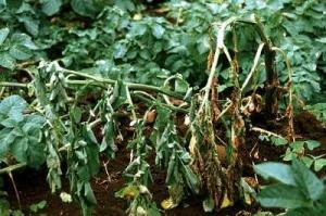
DERZY’S DISEASE

Derzy’s disease is a viral disease also known as Parvovirus disease because of the causative agent. Other names include Goose Plague, Goose Hepatitis, Goose Enteritis, Goose Influenza, Infectious Myocarditis and Ascetic Hepatonephritis. It is a highly contagious disease that affects young geese. The disease has been reported to exist in any part of the world where geese or Muscovy ducks are raised since they are also susceptible to it and can transmit the disease to geese. In its acute form, the disease can result in up to 100 percent mortality rate or it can occur in a more chronic form. If birds are infected during the first week of age, very high losses can occur but if the goslings are 4-5 weeks old or older the mortality rate will be negligible.

Symptoms

For goslings under one week of age the clinical signs are morbidity (anorexia and prostration) and mortality, with deaths occurring in 2-5 days.

Older birds, depending on their level of maternal immunity, will exhibit anorexia, polydipsia, weakness with a reluctance to move, nasal and ocular discharge, swollen and red uropygial glands and eyelids and a profuse white diarrhoea.

Prevention/Treatment

There is no treatment for Derzy’s infection. Adult breeding geese that have been naturally infected with the parvovirus become immune and transfer this passive immunity to their progeny.

This passive immunity will persist in the newly hatched goslings for 2-3 weeks. It is the phenomena of passive immunity being transmitted to the offspring that has led to the development of a recommended vaccination programme.

In its simplest form, all goslings should be vaccinated at about two weeks of age. This assumes that the goslings’ parent flock had been vaccinated which would mean that the goslings? natural passive immunity would protect them until 2-3 weeks of age. 

For birds not designated to be breeders, this single vaccination is sufficient. Birds designated to be breeders should be vaccinated again three weeks before the beginning of lay and three weeks before the beginning of each subsequent lay.

In addition, some practitioners recommend a booster vaccination at peak egg production.

If the parent flock had not been vaccinated which would mean that no passive immunity was passed on to the goslings, the recommendation would be to give serum to the goslings on day one and on day ten to give them passive immunity and to then vaccinate them on day 21.

DUCK VIRUS ENTERITIS

Duck Virus Enteritis (DVE) is an acute, contagious disease caused by a herpes virus that can infect ducks, geese and swans although the incidence of the disease in geese is very low.

DVE can be transmitted directly, by contact between infected and susceptible birds, or indirectly, by contact with a contaminated environment.

Birds that have recovered from DVE are immune to re-infection by the DVE herpes virus.

It should be noted that in Australia a herpes virus has been isolated from a flock of infected geese (with a mortality rate of 97 percent) which was anti-genically distinct from the duck viral enteritis herpes virus.

Symptoms

The symptoms depend on the age and sex of the geese, the stage of infection and the virulence and intensity of the virus exposure.

Lesions of DVE are associated with vascular damage (tissue haemorrhages and free blood in the body cavities), vascular eruptions at various locations on the mucosa surface of the gastrointestinal tract, as well as lesions of lymphoid and other tissues.

Prevention/Treatment

There is no treatment for DVE but vaccines that are effective have been developed.

ERYSIPELAS

Erysipelas is generally an acute, sudden infection of individual geese within the flock. In both young and adult birds it is caused by the bacterium Erysipelothrix rhusiopathiae.

Outbreaks of this disease which are economically significant are uncommon in avian species, with the exception of turkeys, but some cases have been reported for geese. 

Erysipelothrix rhusiopathiae is somewhat unique in that it can infect over 50 animal species and can also infect humans.

In the latter case, the infection usually enters through scratches or puncture wounds and is considered a safety issue for people working with infected animals. Human infections can be treated with antibiotics.

Symptoms

Infected geese will appear depressed, have diarrhoea and die suddenly. Lesions are suggestive of generalised septicaemia.

Treatment
The antibiotics of choice are rapid-acting forms of penicillin that can be administrated together with an erysipelas bacterin. Since the presence of the disease in geese is sporadic, routine immunisation is not generally recommended. However, in areas where the disease is prevalent, and particularly for breeder flocks, vaccination is recommended. Birds that have recovered from acute infections have a high degree of resistance to re-infection.

FLUKES

Flukes (trematodes) are flat, leaf-like parasitic organisms. Over 500 species belonging to 125 genera and 27 families are known to occur in birds. Generally, flukes are not a problem for geese, however, geese with access to natural lake or pond water may become infected. This is because most flukes have an aquatic snail (genus Limnaea) as an intermediate host. The dragon fly (genus Odonata) is the second intermediate host in many cases.

Symptoms

Flukes may invade almost every cavity and all tissue of birds and can show up unexpectedly at a post-mortem. One species of fluke known as the oviduct fluke (Prosthogonimus ovatus), can infect the oviduct which results in flukes appearing in the geese’s eggs.

Prevention/Treatment

The only practical solution is to remove the birds from the source of infection. This can be done if the intermediate host(s) is/are known.

A sample life cycle of flukes (Source: Guy, 1996)
(1) Infected geese excrete fluke eggs in their dropping.
(2) When the conditions are favourable, the eggs hatch, producing a primary larvae.
(3) The larvae mature in an intermediate host (a snail of genus Limnaea).
(4) The intermediate host lays the mature larvae on grass.
(5) After ingesting the larvae by grassing, the geese become re-infected.

FOWL CHOLERA

Fowl Cholera, also known as Pasteurellosis, is a contagious disease affecting all domestic and wild birds. Pasteurella multocida is the causative agent, to which geese are highly susceptible and mortality can be high.

Symptoms

Fowl Cholera usually appears as a septicaemic disease, associated with high morbidity and ortality.

Perhaps the most characteristic aspect of the acute form is the sudden death of birds with the symptoms appearing only a few hours before death.

The chronic form, which can follow the acute form, normally shows as localised infections. The lesions associated with this disease can take several forms, but in most cases the heart, pericardium and air sacs are damaged.

Prevention/Treatment

Fowl Cholera is not a disease of the hatchery nor is it one transmitted through the egg. Rather, infection occurs when the geese are on the farm.

The first step in the control of Fowl Cholera is therefore good sanitary management practices and keeping the geese separate from other birds.

In areas where Fowl Cholera is present either in geese or other species of birds, vaccination of all birds is recommended. In the case of an outbreak, it is possible to treat the birds to stop the spread of the disease, but this must be done quickly.

 LEUCOCYTOZOONOSIS

This is a parasitic disease of birds which affects the blood cells (especially the white blood cells) and the tissues of various internal organs (parasite multiplication occurs in the macrophages of brain, liver, heart, lungs, and spleen).

It is a very uncommon disease in geese but outbreaks of economic significance have been reported. Leucocytozoon simondi is the causative agent in waterfowl and has been reported in 27 species of ducks and geese in North America, Europe and Vietnam.

Symptoms

Leucocytozoon infections are diagnosed by direct microscopic observation and by identification of either the gametocytes (sexual stage of the parasite) in stained blood samples or of the schizonts (stage of massive multiplication) in tissue sections.

Prevention/Treatment 

Treatment of leucocytozoonosis with drugs has, in general, limited success and no effective treatment has been found for Leucocytozoon simondi.

Control methods require the elimination of the insect carriers that include various species of diptera (simuliid flies and culicoid midges) that live near streams.

LISTERIOSIS

Listeriosis is not a common disease of geese but some instances have been reported in temperate areas of the world.

This is probably due to the fact that, in temperate climates, Listeria monocytogenus (the causative agent) is found in both faeces and soil. Also, it is in these areas that many geese are kept on pasture and therefore are exposed to the organism.

Symptoms

The symptoms are septicaemia with necrotic areas in the liver and heart. Encephalitis has been reported in young geese. Infected birds appear emaciated with diarrhoea.

Prevention/Treatment

Prevention depends on eliminating the source of infection. As the organism is resistant to most commonly used antibiotics, high levels of tetracyclines are usually recommended for treatment.

MYCOPLASMA INFECTIONS

Mycoplasma infections, also know as Pleuro-Pneumonia. Like Organisms or PPLO, can cause relatively serious problems in geese.

These organisms have an intermediary structure between that of bacteria and viruses. At least three species of Mycoplasma (Mycoplasma anseris, Mycoplasma claucale and Strain 1220) have been isolated in geese.

In recent years the prevalence of Mycoplasma infections in geese in a number of areas appears to have increased. This is most notable when birds are managed under intensive conditions.

Symptoms

The main problem of Mycoplasma infections is that in breeder flocks it results in reduced egg production and lower fertility.

There is necrosis of the phallus (Venereal Disease) which can cause a severe drop in fertility.

In young goslings Mycoplasma infection results in reduced growth, and respiratory and air sac infections. For young geese the common source of Mycoplasma infection is from the hatching egg.

Prevention/Treatment

The most important aspect of a Mycoplasma control programme is to ensure that the grandparent and parent stocks are Mycoplasma-free so that goslings from these flocks are not infected.

Treatment of eggs from an infected flock is achieved by dipping the eggs in a tylosin solution before the eggs are incubated. Infected goslings can be treated by adding either tetracycline or tylosin to their drinking water.

MYCOSIS OF THE DIGESTIVE TRACT

Mycosis of the digestive tract, caused by Candida albicans, can occur frequently in some classes of poultry but not in geese.

An exception is force-fed birds, where inflammation of the oesophagus may be caused by the insertion of the corn dispenser. This inflammation can then provide a port of entry for Candida albicans.

Symptoms

The symptoms are not particularly characteristic but infected birds show unsatisfactory growth, are stunted, listless and have ruffled feathers. Lesions occur most frequently in the crop and are characterised by a whitish deposit.

Prevention/Treatment

Since unhygienic and overcrowded conditions are conducive to Candida albicans infections, the first step is to eliminate these.

The addition of copper sulphate to the drinking water has had variable results in treating chickens and geese.

Sodium bicarbonate in the drinking water increases the pH in the crop and creates an unfavourable condition for the organism as it likes an acid environment. Addition of either Nystatin or Amphotericin to the feed has been reported to be effective.

 MYCOTOXICOSES

Mycotoxicoses is a disease caused by exposure to mycotoxins, and the most prevalent source of mycotoxin contamination for geese is mouldy feedstuffs.

Diagnosis of Mycotoxicoses can be very complex since hundreds of mycotoxins have been identified.

However, knowing what the geese are being fed, the source, the symptoms the geese are exhibiting and whether or not other livestock or poultry being fed the same feedstuffs are showing similar symptoms, will allow diagnosis of the problem and identification of the source(s) of the mycotoxin.

In tropical countries where aflatoxins are very common, their origin is connected with the development of genus Aspergillus flavus and Aspergillus parasiticus growing mainly on peanuts but also on soybeans, copra, rice bran and corn.

According to the literature, alflatoxins may cause slow growth, a drop in egg production and feather loss for all species of waterfowl, although geese are among the less sensitive.

The genus Fusarium produces numerous toxins injurious to geese, and these have been found in corn, sorghum, barley, sunflower seed, oats, mixed feed and brewers’ grains.

Fusarium mycotoxin production thrives in conditions of high humidity and a temperature of 6-24degC.

In temperate climates it is therefore essential that grains be harvested early before the cool-humid conditions of fall arrive as these are conducive to mycotoxin production.

Symptoms

T-2 toxin is one of the most common Fusarium toxins and, depending on the level of contamination, will cause feed refusal, reduced activity, increased water consumption, reduced egg production and reduced hatch.

There are reports that exposure of young geese to T-2 toxin has resulted in the geese dying within two days.

Another Fusarium toxin to which geese are very sensitive is zearalenone which can not only result in an immediate drop in fertility but can also permanently damage the testes of the gander.

Prevention/Treatment

Treatment is to remove the contaminated feedstuff immediately and provide the geese with fresh, uncontaminated feed. The best prevention is to ensure that all purchased feedstuffs are mycotoxinfree.

NECROTIC ENTERITIS

Necrotic enteritis is caused by Clostridium perfringens and has been reported to occur in geese although the incidence of the disease does not appear to be high. Clostridium perfringens can be found in soil, faeces, dust, litter and contaminated feed.

Symptoms

The clinical signs of Necrotic enteritis are severe depression, decreased appetite, reluctance to move, diarrhoea and ruffled feathers. Sick birds may die quickly due to enterotoxemia and necrosis of the small intestine.

Prevention/Treatment

Prevention is the rule. Many birds have natural populations of Clostridium perfringens in their caeca, but rarely in the small intestine. Stress or any irritant to the digestive tract can provide the stimulus for this genus to appear and multiply in the small intestine and should be avoided. If the disease appears, a number of antibiotics have been found to be effective which include lincomycin, bacitracin, oxytetracycline, penicillin, tylosin, virginiamycin, avoparcin and nitrovin.

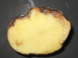
NEPHRITIC HEMORRHAGIC ENTERITIS

Nephritic hemorrhagic enteritis is a disease that is currently quite prevalent in the south western region of France and is often referred to simply as NEHO. It can infect geese from 4-20 weeks of age and causes mortality rates from 30-100 percent. The causes of this disease are not well understood but it seems to be primarily poor management. An excess of protein in the feed or any sudden change in the diet of the birds can also bring it on, as can poor quality drinking water and parasite infections.

Symptoms

When suffering from this disease, geese are often unsteady on their feet, have difficulty getting up and have erratic movements.

These symptoms are accompanied by diarrhoea and trembling and death usually follows shortly afterwards.

The characteristic lesions are urates and haemorrhaging in the kidneys, an exaggerated sub-cutaneous swelling and the presence of intestinal parasites.

Prevention/Treatment

The first measures to take are good management prevention practices such as controlling parasites and ensuring that the geese have a balanced ration. For outbreaks of the disease, good results can be obtained by injecting homologous serum.

Also available are renal tonics and liver detoxicants, both of which can help relieve the symptoms. Due to a lack of knowledge of the disease, no vaccine has yet been developed.

NEWCASTLE DISEASE

The Newcastle Disease Virus is of the genus Paramyxoviruses which has been isolated from geese. Clinical signs are the exception rather than the rule, but when present, consist of greenish diarrhoea and, occasionally, disorders of the central nervous system.

In many cases, geese may be infected without showing any clinical symptoms, yet they can be carriers for a prolonged period.

Usually geese are not vaccinated since Newcastle disease is not generally a problem for them.

PARATYPHOID

Paratyphoid, or salmonellosis, is an important disease in geese with young birds, generally under six weeks of age, being the most susceptible. In addition, the concern regarding salmonella infection in humans and the demand for salmonella-free poultry products has increased the awareness of this disease and resulted in various monitoring programmes being undertaken in many countries.

Over 2 000 types of salmonella organisms have been isolated from various species of fowl worldwide. Generally, the salmonella serotypes isolated from poultry are more characteristic of the region than the species of poultry.

Paratyphoid is easily spread through contact with either infected birds, their faeces or through infected equipment, particularly that used for hatching and brooding. It now appears that salmonella is spread by salmonella entering the egg both in vivo before it is laid and by penetrating the egg after it is laid.

In both cases it can multiply in the egg. For this reason, the importance of collecting eggs frequently before they get dirty, and cleaning and fumigating them as soon as possible, cannot be over emphasised.

Symptoms

Geese with Paratyphoid will usually be less than six weeks of age, tend to stand in one position, with their heads lowered, eyes closed, wings dropping and feathers ruffled. Sick birds will also exhibit marked anorexia, increased water consumption, watery diarrhoea, pasty vent and a tendency to huddle close to the heat.

Prevention/Treatment

The first step in the control of Paratyphoid is to remove all the possible sources of salmonella. This requires excellent management and sanitation of the breeders, the hatching process and the rearing of the goslings.

The cleanliness of the hatching eggs is perhaps the most important single aspect in the control of Paratyphoid, especially the fumigation of eggs immediately after laying.

Rodent control is also very important. A number of sulphonamides, antibiotics and nitrofurans have been recommended in the treatment of paratyphoid. In addition, furazolidone and injectable gentamicin and spectinomycin can be used.

The final diagnosis of Paratyphoid depends on isolation and identification of the causative organism. This will help determine which drugs are best suited to treat a particular outbreak.

RIEMERELLA ANATIPESTIFER INFECTION

Riemerella anatipestifer infection is a contagious disease affecting domestic geese, ducks and various other birds which means that infections in geese can originate from other species.

Symptoms

The common symptoms are ocular and nasal discharges, mild coughing and sneezing, greenish diarrhoea, uncoordinated movement, tremor of the neck and head and coma.

Geese that recover from the disease are resistant to subsequent infection.

Prevention /Treatment

The sulphonamides and antibiotics as listed under Fowl Cholera for the control of Pasteurella multocida are usually effective against Riemerella anatipestifer. Vaccines have been developed but they have been used primarily with ducks although they can be expected to prevent the disease in geese as well.

 PSEUDOTUBERCULOSIS

Pseudotuberculosis caused by Yersinia pseudotuberculosis has been reported in a large number of avian species, including geese. It is not, however, a common disease in geese.

Symptoms

The disease is characterised by an acute septicaemia and infected birds have difficulty breathing and are weak, with dull and ruffled feathers and diarrhoea.

A definite diagnosis requires isolation and identification of the causative agent.

Prevention/Treatment

Due to the low incidence of the disease, there is very little information available but chloramphenicol, streptomycin and tetracycline have been effectively used in some species.

RETICULOENDOTHELIOSIS

Reticuloendotheliosis refers to a group of syndromes caused by the retroviruses of the REV group. The disease occurs in a wide variety of domestic poultry but is rare in geese.

It is sometime called the Runting Disease because it is characterised by poor growth and abnormal feathering.

In geese, viruses have been isolated from tumours of the spleen, liver, pancreas and intestines. No vaccine has been developed for this disease because the incidence and economic importance of the disease is very low.
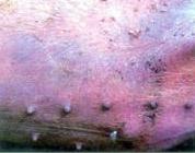
SPIROCHETOSIS

Spirochetosis in avian species is caused by Borrelia anserina and is tick-borne. Spirochetosis was first described in 1891 as a severe septicaemic disease of geese in Russia but it is now found worldwide, especially in the tropical and subtropical areas where fowl ticks (genus Argas) are common. However, even in these areas the incidence of the disease is low.

Symptoms 

Morbidity and mortality are highly variable, ranging from 1-2 percent up to 100 percent.

Lowest rates occur when the birds have previously been exposed to Borrelia anserina and have developed immunity. Larval ticks or puncture haemorrhages from tick bites on the birds, or ticks in the birds’ environment are indicative of the disease.

Prevention/Treatment

In areas where Spirochetosis is prevalent, vaccination is the control method of choice.

Female geese that have acquired immunity, either through natural exposure or through vaccination, are capable of passing on passive immunity to their offspring which will protect them for 5-6 weeks post hatching.

When an outbreak occurs, the treatment of choice is usually antibiotics. Borrelia anserina is sensitive to most antibiotics including penicillin, chloramphenicol, kanamycin, streptomycin, tylosin and tetracyclines.

STAPHYLOCOCCOSIS

All avian species are susceptible to staphylococcal infections though geese do not appear to be affected to any great degree.

If and when they are infected, it is generally as a secondary infection but even this is rare.

Staphylococcus aureus is the most common infection in birds. One of the major concerns is that staphylococcus infections can be transmitted from birds to humans.

This has been observed among both slaughterhouse workers and people performing autopsies.
The most frequent sites of infection in poultry are bones, tendon sheaths and leg joints but infections may occur elsewhere.

Prevention/Treatment

Staphylococcus infections can be treated with antibiotics. Penicillin, streptomycin, tetracycline, erythromycin, novobiocin, sulphonamides, linomycin and spectinomycin have been used successfully.

STREPTOCOCCOSIS

There are a number of species of streptococcus that infect birds. However, to date, streptococcus infections in geese are very rare although Streptococcus mutans, a common bacterium of the human oral cavity, has been identified as a cause of septicaemia and mortality in geese.

Symptoms

In its acute form, the clinical signs of Streptococcosis are related to septicaemia, depression, lethargy, diarrhoea and head tremors, although often the birds are just found dead. In the chronic form, depression, loss of weight, lameness and head tremors may be observed.

Prevention/Treatment

Prevention and control require reducing stress and following proper sanitation practices. Treatment includes the use of either antibiotics such as penicillin, erythromycin, tetracycline or nitrofurans.

TRICHOMONIASIS

This is a protozoan disease that infects mostly mature geese in breeder flocks. The causative agent in geese is Trichomonas anseris while for other classes of poultry it is Trichomonas gallinae. These organisms are transmitted from bird to bird through the water and, to a lesser degree, through the feed.

Symptoms

The infection in geese is mainly in the lower digestive tract and the first symptoms are reduced reproductive performance and weight loss. The droppings can be monitored for the protozoan although an autopsy (with heavy infections mortality can be high) will generally not yield the protozoa as they disappear quickly.

Prevention/Treatment

If the disease has not spread throughout the flock, any sick birds that can be identified should be isolated. Nitrofurazon, metronidazole and dimetridazole are effective in treating the disease.

VENEREAL DISEASES

Bacteria, especially Neisseria, Mycoplasma, and Candida albicans have been associated with a venereal disease in ganders although it now seems that Mycoplasma are the primary infective agents. 

Symptoms

Initially, the base of the phallus becomes swollen and inflamed with the infection extending to the cloaca.

Later, there is necrosis, ulceration and eventually considerable scarring, making reproduction impossible. The disease spreads throughout the flock very rapidly.

Prevention/Treatment

The onset of the disease has, in some cases, been associated with a high density of ganders that has led to fighting, resulting in the phallus of some ganders being injured and becoming infected.

The infection then spreads through the flock via the females. When infected, the females exhibit symptoms such as airsaculitis, peritonitis, and salpingitis.

The first control measure to take is good management of the breeder flock. Because of the principle involvement of Mycoplasma, some veterinarians view the disease as a component of Mycoplasma infections rather than as a separate disease.

Treatment is therefore with antibiotics effective against mycoplasma such as tylosin, tetracycline, chlortetracycline, linomycin, oxytetracycline, spectinomycin, spinomycin and tiamulin. Sensitivity tests should be conducted to select the appropriate antibiotic.


Common Pests and Parasites in Pigs

Credits:Biovision-Infonet

Parasitic diseases

Parasites are divided into external and internal parasites.

Internal parasites (Worms)

Worms are one of the most serious threats to pig keeping. There are more than 30 types affecting the intestines of pigs. The most important are the intestinal roundworm, the lung worm, the whip worm and the tape worm.

1) Roundworm

Roundworms live in the gut and take food from the pig. The pig can therefore become thin. 
Symptoms: 

  • Anorexia (loss of appetite) in advanced stages.
  • Anaemia (loss of condition). 
  • Weight loss in later stages. 
  • When the worms die suddenly after treatment, they can block the gut and cause sudden death. 

Prevention:

  • Control with medicine in the food is useful and provide clean and dry pens.
  • Separation of young ones from adults. 
  • Washing sows before farrowing.
  • If pigs are allowed outside, rotational grazing and periodic resting of pastures to allow disinfection. 

Treatment:

  • Periodic deworming
  • Herbal treatment: herbs such as Moringa are considered to be antihelmintic ( able to kill intestinal worms)

2) Whipworm

Symptoms:

  • Anaemia
  • Haemorrhagic diarrhoea causing anaemia 
  • Weight loss

Prevention and treatment: 

  • Deworming

3) Lungworm

Symptoms:

  • Coughing

Prevention

  • Deworming
  • Clean pens 

Treatment

  • Riperol (or Levamisole) injections are used in treatment, but are may be difficult to obtain in many places.

4) Tapeworm (measles)

Pork measles is caused by tapeworms which live in the muscles of pigs. They do not usually affect the pig, but can lead to pain and the pig may find it difficult to move around. When people eat undercooked measly pork, the worms develop inside the people, and can make them very sick.
Symptoms:

  • Poor growth
  • Rough grey hair coat
  • Swollen belly
  • Emaciation
  • Anaemia

Prevention

  • Prevent the pigs from wandering about where they can feed on human faeces
  • Make sure that people working with pigs use toilets (hygiene and sanitation)
  • Deworming

External parasites

External parasites include mange, lies and myiasis (see below).

Mange

It is caused by small parasites called mites that live in the skin. They provoke severe itching and irritation.
Symptoms

  • The pig becomes itchy, and scratches and rubs against the walls of the sty and other objects with the skin between the legs, around the eyes, ears and neck being principally affected.
  • The coat looks dull, and there are bare patches, heavy crusts, and lines on the body that look like ribs 
  • Restlessness and itching which can be very severe 
  • Red pimples on skin, which turn into crusts and scabs. Later the skin looks very rough, is thickened and covered with flakes scratching. Skin may show red spots or bite wounds
  • Thick skin and rough hair coat
  • Anaemia in severe cases especially in piglets
  • Death in severe cases

Prevention:

  • Wash the sow before farrowing at least twice at a one week interval. 
  • Inspect and examine gilts carefully upon entering the farm, and treat them if necessary.
  • Examine the animals before serving and treat them if relevant. 
  • Wash all pigs at the beginning of fattening if mange is already a problem. 
  • General cleanliness.
Crusts seen within the ear of a pig due to mange infestation
(c) S. Gikonyo, Kenya

Treatment:

  • Remove scales and dirt with soap and water and a stiff brush. 
  • Afterwards the pig should be washed with organophosphate compounds. Repeat this treatment several times
  • Ivermectin injection is a very effective treatment against mange and all other parasites. Spraying the animals with cattle dip also kills many parasites on the skin
  • Herbal treatment: Smearing with coconut oil can be an effective control in cases of light contamination


Lice 

These are blood suckers that also cause irritation of the skin. 

Symptoms

  • Itching 
  • Skin may show red spots or bite wounds. 
  • Thick skin and rough hair coat. 
  • Anaemia in severe cases especially in piglets. 

Prevention:

  • General cleanliness
  • Examine piglets before putting them in fattening house, and treat if relevant. Fatteners don’t need to be treated
  • Examine gilts before first service, and treat if relevant
  • Examine boars twice a year, and treat if relevant
  • Examine new stock on arrival and seven days later. Piglets below three weeks should not be treated. Treat if it is found relevant.

Treatment:

  • The insecticide benzene hexachloride is a very effective remedy (0.1 – 0.25% solution) against lice. Treatment should be repeated after 7 days since the insecticide only kills adults and not eggs. 
  • Lice can be kept away to a certain extent by planting a pole sturdily in the ground at an angle of 45deg with an old sack wound around it immersed in crude oil or used motor-oil.

Myiasis 

Disease caused by flies, which lay eggs in the wounds. The eggs hatch into larvae which live and feed on the flesh and fall off when they mature, creating more wounds.
Symptoms

  • Infected wounds look very wet and dirty and the edges can be covered with a grey mass which are the eggs of flies. 
  • Later on, larva can be seen as screw-shaped pinkish worms crawling through the wound. 
  • The pigs show annoyance and try to find a shady place. 
  • Death may occur.

Prevention and treatment 

  • Clean the wound daily with water and disinfectant then apply insecticide on wound to cover edges too. 
  • Best working insecticides are the long-acting ones e.g. diazinon or supona. Whenever using insecticides, check with the organic standards whether it is allowed, and minimize it as much as possible, and seek alternatives.
  • Good mange control program to prevent wounds smooth walls and floors.
  • Do not use sharp objects.
  • Avoid overcrowding to reduce fighting.
  • Tail docking, ears notching and castration should be handled properly and the umbilical cord disinfected.

General recommendation

1) Deworming 

  • Boars – every 6 months
  • Sows – 2 weeks before farrowing and after weaning
  • Piglets – 1 week after weaning
  • Fatteners – 1 week after weaning and 3 months latter 
  • Gilts – 1 week after weaning, at 3 months and at 7 months of age and at least 2 weeks before service.

2) Control of Lice, Fleas, Mange 

  • Scrub sows with Gamma BHC insecticides four days before farrowing. 
  • Clean and scrub the farrowing pens before use.