Calf Feeding

Dr.iCow’s Diary

Date: 09.02.2020

Dear Dr.iCow,

My cow has given birth. Is it good for the calf to suck the mother?

From: Teresia, County: Machakos, Kenya.

Discussion:

Dear Teresia,

Congratulations! Please let the calf suck for the next 48 hours.

At birth and on standing allow the calf to suck colostrum from the mother. You may start milking a cow immediately after calving if the udder or teats conformation prevent easy first sucking and provide the new born calf with colostrum using a milk feeding bottle. If the calf can suckle without difficult let the calf suck the colostrum and bond with the mother for a maximum of 2 days. The colostrum is very important for the calf because it contains high levels of antibodies and vitamins A and D the calf needs to prevent diseases caused by micro-organisms on the farm.

A calf is born with few antibodies of its own and has immature immune system which is not capable of producing antibodies for some weeks. Colostrum is highest absorbed from birth to 4 hours best within 12 hours good within 24 hours and very low after 24 hours. It is the first meal for the calf and is rich in nutrients as it is high in energy, proteins and vitamins than normal milk. Ensure the calf gets plenty colostrum within the first 6 hours after birth. In the first 20 minutes the calf would need about 2 litres of colostrum and 2 litres after 6 hours. If the calf is sucking allow it to suckle for at least 20 minutes. Please observe high level of hygienic standards.

The first milk or colostrum should be fed to the calf. A calf’s digestive system start to change as soon as it born and; at birth it has the greatest ability to absorb antibodies from the colostrum. Calves of dairy cows are separated from their mothers within the first 24 hours after birth. The calves need close monitoring and extra attention should be provided as early separation cause stress.

Thank you

From your friend and advisor,

Dr.iCow
Cassava Anthracnose

(Glomerella manihotis)

Credits: Biovision-Infonet

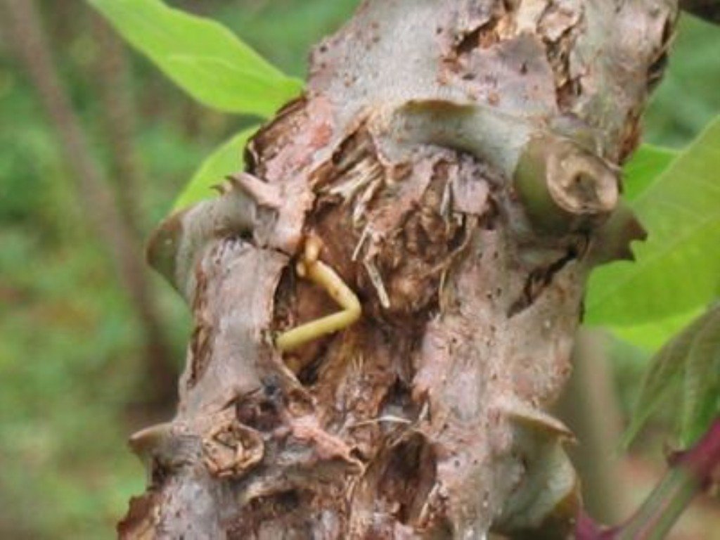
Severe stem infection by cassava anthracnose
(c) IITA, 2010
Cancers of cassava anthracnose disease (Glomerella manihotis) on stem
(c) www.iita.org

Initial symptoms of the disease are oval lesions (“sores”) on young stems. On older stems, raised fibrous lesions develop that eventually become sunken.

What to do:

  • It is not an economically important disease in most cassava growing countries and it does not warrant any intervention.

Cassava Brown Streak Virus Disease

(Potyvirus – Potyviridae)

Credits: Biovision-Infonet

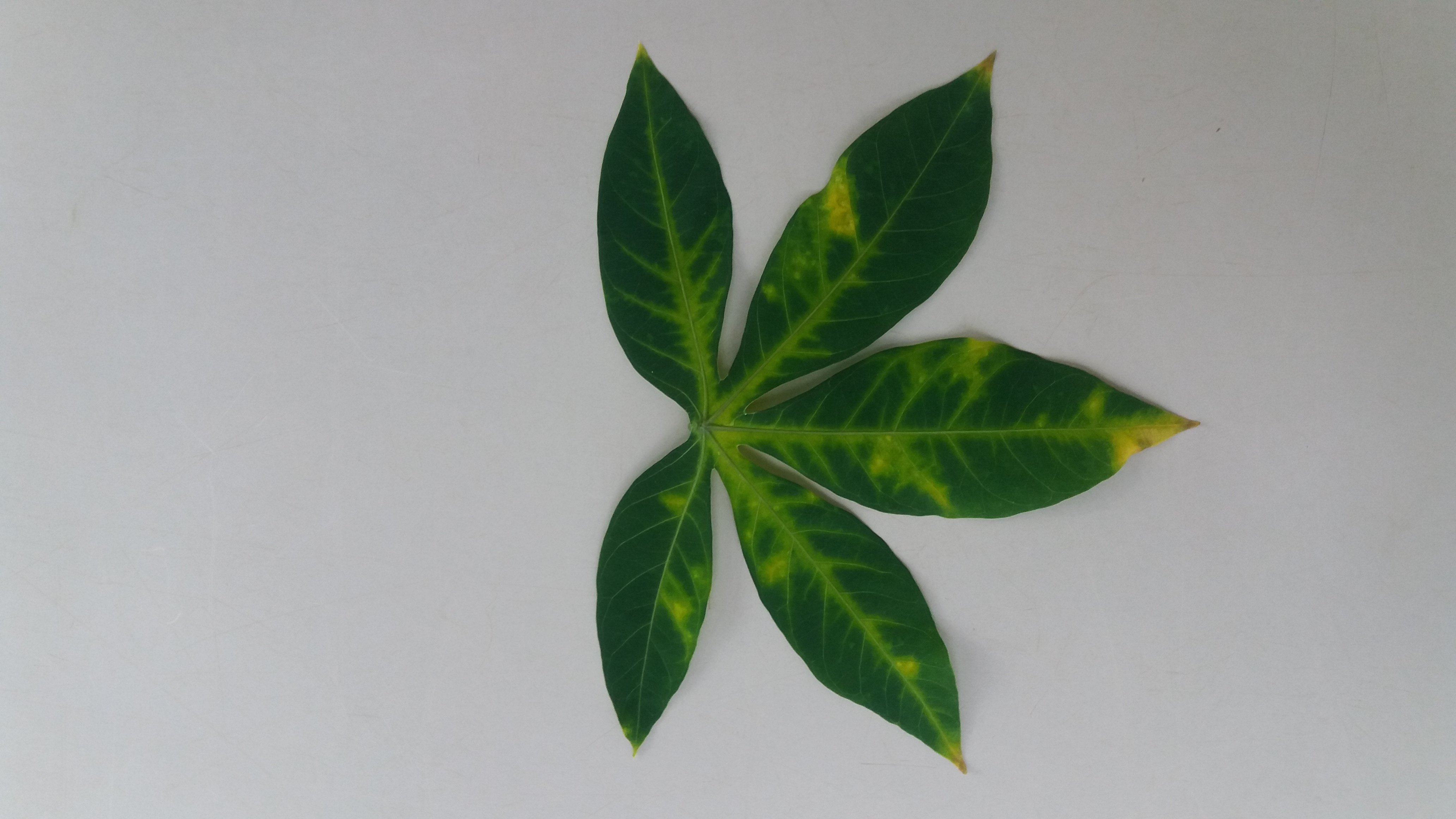
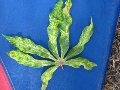
Cassava brown streak virus symptoms on plant. Notice chlorosis in between veins.
(c) Emily Masinde, 2018
Cassava brown streak virus symptoms on plant. Notice chlorosis along leaf veins.
(c) Emily Masinde, 2018
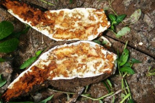
Cassava roots completely destroyed by Cassava Brown Streak Disease. This disease renders cassava roots unfit for consumption and use.
(c) IITA, 2010

It is particularly serious in coastal areas of Kenya, Zanzibar, Mozambique and Tanzania and lakeshore region of Malawi and in Uganda and is a threat to the whole of sub-Saharan Africa.

The virus is vectored by whiteflies (Bemisia spp.) and also transmitted through infected cuttings. Symptoms include yellowing (leaf chlorosis) and brown streaks in the stem bark (cortex).

Infected tubers have brown streaks (root necrosis) (Field Crops Technical Handbook, MoA, Kenya). It’s a stealth virus, which destroys everything in the field. The leaves may appear healthy even when the roots have rotted away.

What to do:

  • Use diseased-free cuttings.
  • Use tolerant/resistant varieties (e.g. “5543/156”, “TMS 30572”)
  • Remove diseased plants from the field.

Feeds contaminated by Mycotoxins

Dr.iCow’s Diary

Date: 07.02.2020

Dear Dr.iCow,

Ng’ombe yangu inahara damu.

Translation: My cow is diarrheaing blood

From: Anne, County: Uasin Gishu, Kenya.

Discussion:

The cow is eating normally and she is not showing any signs of illness except there is blood in the dung. Anne had some spoilt maize grains crushed and made some maize by-products concentrates from the spoilt maize which she has been feeding the cow. The grains and the maize by-product concentrates could be containing some mycotoxins which are toxic metabolic by-products of fungal growth on grains. These mycotoxins can seriously impair an animal’s health and productivity. Aflatoxins B1 is the most potent compared to the other mycotoxins. The mycotoxins may be the cause of the presence of blood in the cow’s dung. 

Dear Anne,

Presence of blood in the cow’s dung could be due to infections like coccidiosis, mycotoxins and salmonellosis. Mycotoxins are toxic metabolic by-products of fungal growth on grains. High moisture content of grains can lead to fungal growth and toxins production before or after grain harvest. The concentrates you have made for the cow using spoilt maize could be containing mycotoxins. It is advisable to withdraw and stop feeding the cow with this concentrate feed you have made. If blood continue to appear in the cow’s dung or in the manure call a vet immediately.

Aflatoxins are poisonous by-products of the fungal growth – the mold fungus, Aspergillus. Aflatoxicosis is a disease caused by the consumption of aflatoxins, mold metabolites produced by strains of Aspergillus flavus and Aspergillus parasitisus. Contaminated grains and grains by-products are the most common sources of aflatoxins in the country.

The common aflatoxins are B1, B2, G1 and G2. Cattle and young animals are most susceptible to aflatoxicosis. Signs of chronic aflatoxicosis are poor feeding, decreased milk yields, reduced feed conversion efficiency and poor growth rate. Others are weight loss, mild dirrhoea, rough hair coat, listlessness and anemia. It can cause abnormal heat cycle, impair reproduction and immunity and in dairy cows aflatoxins metabolites can appear in the milk before these signs develop.

Animal feeds and feed ingredients should be purchased from reliable feed companies and their storage should be at proper moisture levels.

Thank you

From your friend and advisor,

Dr.iCow
Brown Leaf Spot

(Cercosporidium henningsii)

Credits: Biovision-Infonet

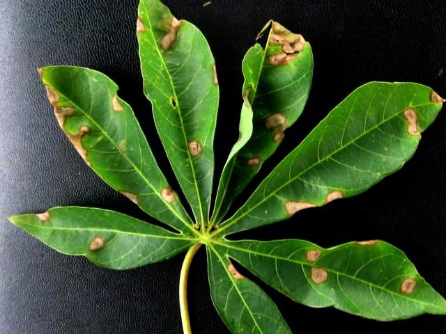
Cassava brown leaf spot (Cercosporidium henningsii)
(c) A.A.Seif, icipe

Symptoms are restricted to older leaves. Brownish round spots with definite borders appear on the upper leaf surface.

On the lower leaf surface, they are brownish-grey in colour. Infected leaves later become yellow and eventually drop. In wet areas the disease may cause a yield reduction of up to 20%.

What to do:

  • Though the disease is widespread in most cassava growing countries, it is not an economically important disease problem and it does not warrant any intervention.

Cassava Bacterial Blight

(Xanthomonas campestris pv. manihotis)

Credits: Biovision-Infonet

Cassava bacterial blight (Xanthomonas campestris pv. manihotis). Angular leaf spots, sometimes with yellow haloes, rapidly expanding, leading to necrosis and leaf fall.
(c) Grahame Jackson (Courtesy of EcoPort)

It is a major constraint to cassava cultivation in Africa. Infected leaves show localised, angular, water-soaked areas. Under severe disease attack heavy defoliation occurs, leaving bare stems, referred to as “candle sticks”. Since the disease is systemic, infected stems and roots show brownish discolouration.

During periods of high humidity, bacterial exudation (appears as gum) can readily be observed on the lower leaf surfaces of infected leaves and on the petioles and stems. The disease is favoured by wet conditions.

This disease is primarily spread by infected cuttings. It can also be mechanically transmitted by raindrops, use of contaminated farm tools (e.g. knives), chewing insects (e.g. grasshoppers) and movement of man and animals through plantations, especially during or after rain.

Yield loss due to the disease may range from 20 to 100% depending on variety, bacterial strain and environmental conditions.

What to do:

  • Use clean planting material. This can reduce disease incidence in areas where cassava bacterial blight is already widespread.
  • In cases of sporadic occurrence of the disease, collect cuttings only from healthy plants and from the most lignified portion of the stem, up to 1 m from the base. Check visually the cuttings for vascular browning. Disinfect tools regularly.
  • Intercrop cassava with maize or melon. This been reported to reduce cassava bacterial blight significantly.
  • Practise crop rotation and fallowing. These practices proved very successful when the new crop was planted with uninfected cuttings. Rotation or fallowing should last at least 1 rainy season.
  • Remove and burn all infected plant debris and weeds. Alternatively plough them into the soil.

Lumpy Skin Disease

Dr.iCow’s Diary

Date: 06.02.2020

Dear Dr.iCow,

How to treat Lumpy skin disease. Which drugs should I use?

From: Mr. Odingo Benedict,County: Homa Bay, Kenya.

Discussion:

The cow has nodules all over the skin, poor feeding and very weak. The vet had treated the cow with some injections but still is not showing improvement.

Dear Mr. Odingo,

Lumpy skin disease – LSD is a viral disease that affects cattle. It is a very devastating disease. Signs associated with the disease are high fever of up to 41ºC, swollen lymph nodes, development of large firm nodules of about 2 to 5 centimeters in diameter and are found all over the animal body, the nodules may spread to other parts of the body including respiratory and gastrointestinal tract and the disease can be fatal.

It is a notifiable disease. It is very good you reported the case to the veterinary officer. There is no treatment of the disease. The vet gave an antibiotic to prevent pneumonia and skin infections, and may also give vitamins and minerals supplements injection like Catosal as the sick animal is having suppressed feed intake and disease stress.

Other supportive treatment include wound care using products like tetracycline spray or healing oil on skin lesions. Provide the cow with  adequate and quality forages with fine texture and mixed with legumes like sweet potatoes vines all mixed together, chopped, and molasses may be added to improve the feeds energy content, palatability and taste, give minerals supplements mixed with concentrates and easy  free access to clean fresh drinking water. The disease takes long time to heal and can persist for months. 

Lumpy skin disease is controlled through vaccination, control of animals’ movement and culling of infected ones. Please make sure your cattle and of the neighbours are vaccinated to protect them from LSD and the vaccination is done once in a year by the county veterinary department.

Like all viral infections, there are no specific antiviral drugs for its treatment. Sick animals should be removed from healthy ones and given supportive treatment. The disease is prevented by effective vaccination.

African Cassava Mosaic Disease (ACMD)

Credits: Biovision-Infonet

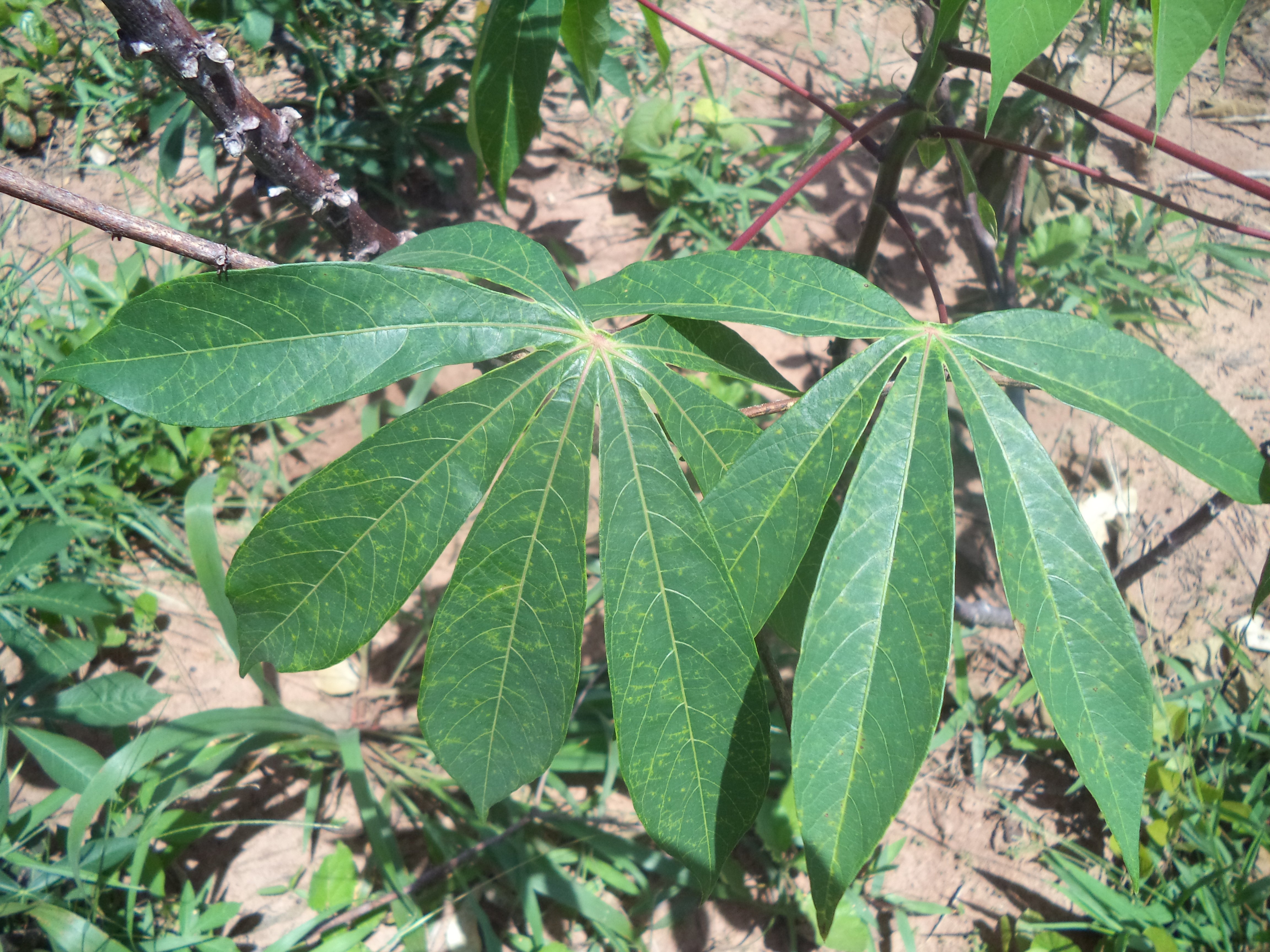
Cassava plant showing severe symptoms of the African Cassava Mosaic Disease (ACMD).
(c) A.A. Seif, icipe
African Cassava Mosaic Virus symptoms
(c) A.A. Seif, icipe

African cassava mosaic disease is one of the most serious and widespread diseases throughout cassava growing areas in Africa, causing yield reductions of up to 90%.

It is spread through infected cuttings and by whiteflies (Bemisia tabaci).

Symptoms occur as characteristic leaf mosaic patterns that affect discrete areas and are determined at an early stage of leaf development.

Symptoms vary from leaf to leaf, shoot to shoot and plant to plant, even of the same variety and virus strain in the same locality. Some leaves situated between affected ones may seem normal and give the appearance of recovery.

What to do:

  • Use disease-free cuttings. If it is not possible to find cassava plants that are completely free from the disease, select cuttings from stem branches instead of from the main stem. Stem cuttings from the branches are more likely to sprout into disease-free plants than stem cuttings from the main stems (James et al, 2000).
  • Resistance to ACMD has been successfully incorporated into high yielding cultivars of acceptable quality through breeding programmes at IITA. Use resistant/tolerant varieties (e.g. “SS 4”, “TMS 60142”, “TMS 30337” and “TMS 30572”)

Quick Search Cassava Pests

Credits: Biovision-Infonet

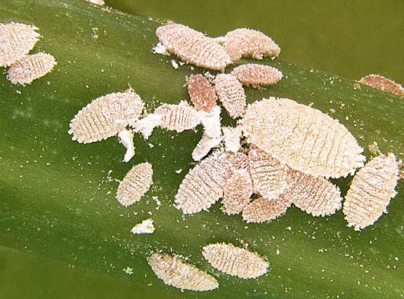
Cassava mealybug

The cassava mealybug is pinkish in colour. Its body is surrounded by very short filaments, and covered with a fine coating of wax. Adults are 0.5 – 1.4mm long.
(c) G. Goergen (Courtesy of EcoPort, www.ecoport.org)
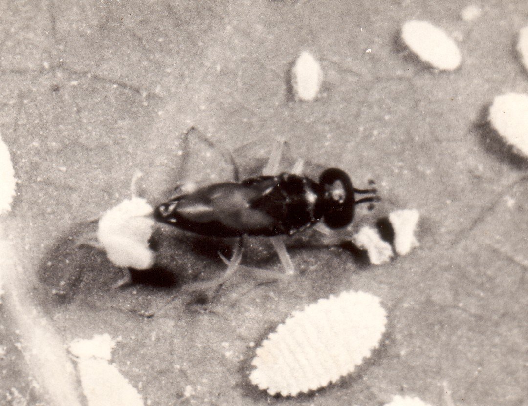
Parasitic wasp of mealybugs
(c) A.M. Varela, icipe
Mealybug infestation on lower leaf surface of cassava plant
(c) A.A. Seif

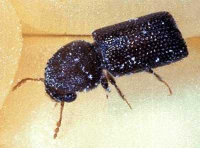
Larger grain borer

Larger grain borer (Prostephanus truncatus). The adult beetle is 3-4.5 mm long.
(c) NRI/MAFF. Reproduced from the Crop Protection Compendium, 2004 Edition. (c) CAB International, Wallingford, UK, 2004

Birds and other vertebrate pests

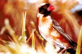
Red-Billed Quelae Bird is one of the most dangerous of all agricultural pests in Africa, and causes food shortages in many countries. The flock breeds at times of abundant rainfall and young are ready to move with the nomadic flock within six weeks,often coinciding with the ripening of grain crops. A nesting colony of Red-billed quelea can extend over hundreds of acres, and a single flock may number millions of birds, moving together in a synchronized fashion. Recent discussions about quelea bird pest control have started to turn towards prediction of breeding based on weather patterns
(c) Courtesy EcoPort (http://www.ecoport.org) : C.Elliott.
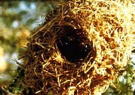
Red-Billed Quelae Bird Nest The nest is made in the form of a pouch with a roof and an entrance near the top. The male is responsible for the construction which is completed by a remarkable technique of weaving strips of grass back and forth to form a mesh-like structure. This is why the quelea bird is called a ‘weaver bird’. If the nest is accepted by a female, she will enter and in due course produce a clutch of three eggs.
(c) Courtesy EcoPort (http://www.ecoport.org) : C.Elliott
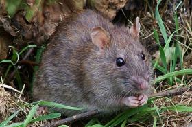
Wild Rat
(c) Reg Mckenna, wikipedia
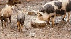
Free-range pig keeping
(c) S. Gikonyo, Kenya
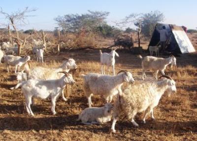
Angora and Galla goats
(c) Anne Bruntse, BioVision

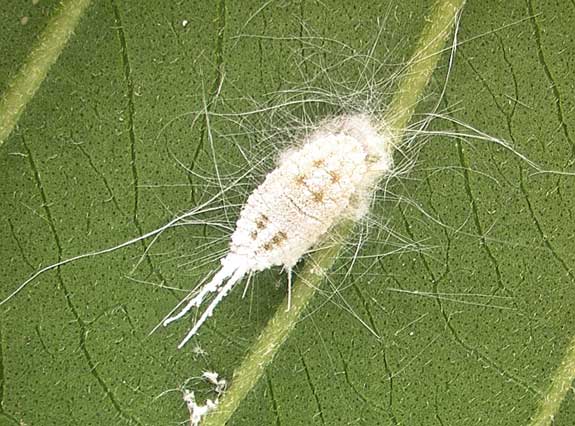
Striped mealybug

Striped mealybug (Ferrisia virgata)
(c) F. Haas, icipe

Cassava green spider mite

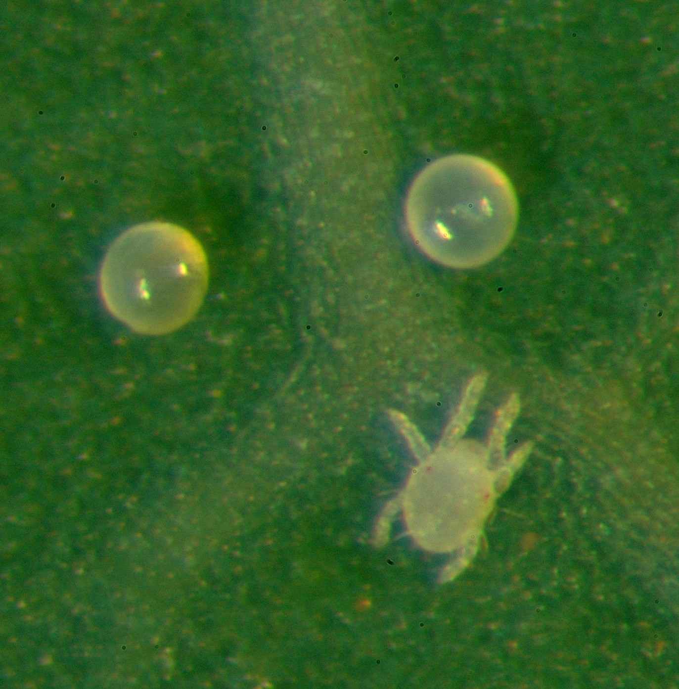
Cassava green mite (Mononychellus tanajoa) and eggs. Real size 0.8 mm, egg 0.2 mm.
(c) F. Haas, icipe
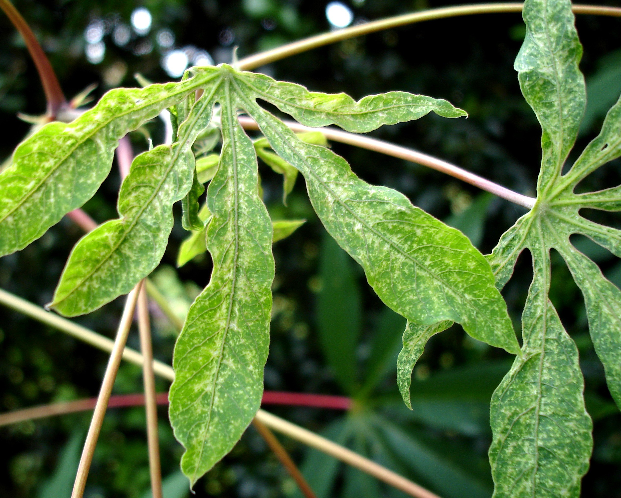
Damage by the cassava green mite (Mononychellus tanajoa)
(c) A. M. Varela, icipe

Red spider mites

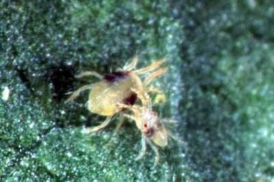
Two-spotted spider mite (Tetranychus urticae) . The adult female is 0.6 mm long. The male is smaller.
(c) Warwick HRI, University of Warwick

Cassava scales

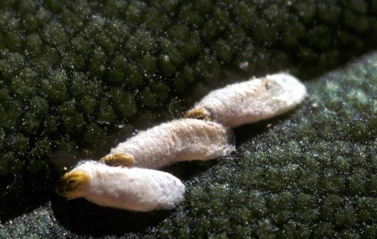
Scale insect. This is not the cassava scale, but an armored scale (related species)
(c) USDA ARS, Bugwood.org

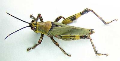
Grasshoppers

Variegated grasshopper (Zonocerus variegatus)
(c) Georg Goergen (Courtesy of EcoPort, www.ecoport.org)

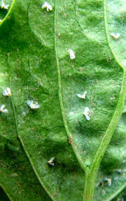
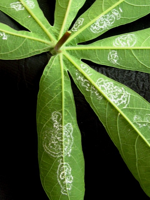
Whiteflies

Whiteflies on chilli leaf. Adults are about 1mm long.
(c) B. Nyambo, A. A. Seif, icipe
Whiteflies egg laying on lower leaf surface of cassava plant
(c) A.A. Seif

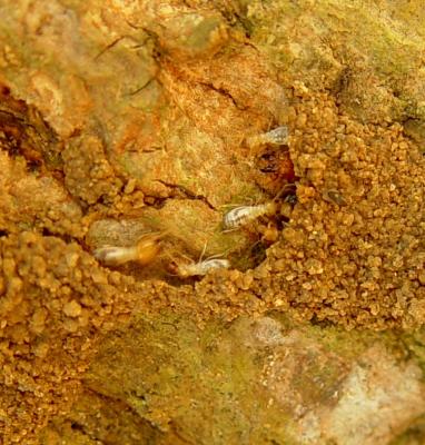
Termites

Close-up termites on mango stem.(Coptotermes formosanus)
(c) A. M. Varela, icipe

Storage pests

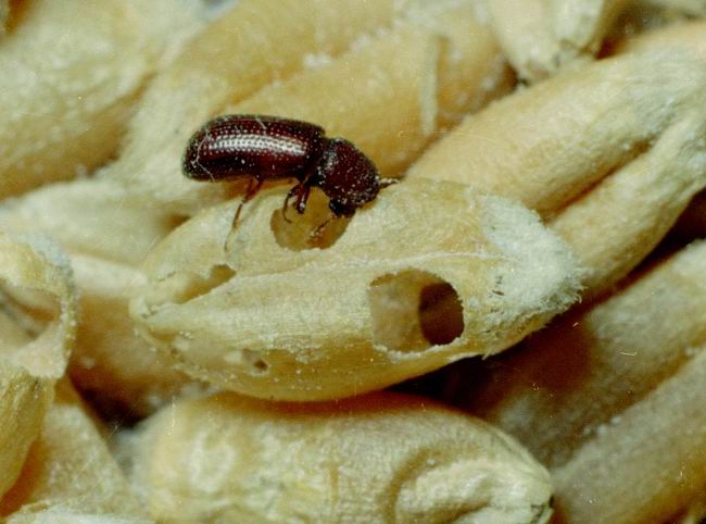
Lesser grain borer (Rhyzopertha dominica). Adults are 2-3 mm in length and reddish-brown in colour (shown on wheat grains).
(c) Clemson University – USDA Cooperative Extension Slide Series, United States, bugwood.org
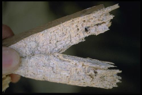
Damage to cassava chips by Larger Grain Borer
(c) GTZ

Dehorning in Cattle

Dr.iCow’s Diary

Date: 05.02.2020

Dear Dr.iCow,

My cow has a problem in the horn of left with a lot of something like mucus. Tell me doctor.

From: Robert Mutuma, County: Meru, Kenya.

Discussion: 

The cow was dehorned in August 2019. Wounds usually heal well with no treatment. There are risks of complications like uncontrolled bleeding, fly contamination and bacterial infections and they do occur.  In this case the dehorning wound healed but now some pus has been coming out on the left horn wound and this could be because of bacterial infection through contamination.

Dear Robert,

The cow with a problem of pus coming from the left horn wound after dehorning is having a complication of bacterial infections. Dehorning wounds usually heal well and quickly. The pus around the left horn following dehorning in August last year shows that the wound did not heal properly and the dehorned site could be septic or contaminated. The cow could be suffering from a condition called suppurative frontal sinusitis. Please call a vet doctor to examine and give treatment which may involve a minor surgical procedure.

Please note, after dehorning it is very important to have a post-operative management so as to prevent bacterial contamination. Poor management of wounds can result in very serious complications. Dehorning is a delicate procedure and should be performed by qualified veterinary professionals.

It is advisable to dehorn animals at young age at least before they attain the age of 2 months when horn buds become attached to the frontal bone of the skull.

Dehorning is part of cattle management. Dehorning has the benefit of having a herd free from damage like severe injuries and bruising caused by horns. There are various choices of dehorning methods like; chemical dehorning hot-iron dehorning and surgical disbudding. Disbudding and dehorning are surgical procedures. Calves and cattle require observation and aftercare following the procedure. Always consult a veterinary doctor.

Thank you

From your friend and advisor,

Dr.iCow