Change of hair coat in cows

Dr.iCow’s Diary

Date: 13.02.2020

Dear Dr.iCow,

Ni Sylvester kutoka Bomet. Kuna ng’ombe ambaye alikuwa mweupe (pepepe) na saa hii ni nyeusi (nyeusi tititi), shida ni nini?

My cow had a white hair coat and it has now changed to black, what could be the problem?

From: Sylvester, County: Bomet, Kenya.

Discussion:

The cow had a hair coat of white colour and now has changed to black. He has tried to give vitamins but there is no change but continue to get darker. Sylvester has different explanations on the colour change which is unusual and says he had another case similar to the current one and which responded to multivitamin treatment. Deficiency of some trace minerals like copper may precipitate the colour change.

Dear Sylvester,

The colour change of the cow’s hair coat could be due to nutritional and minerals deficiencies like copper.  Other causes are chronic disease, mycotoxins, and liver damage by some plants toxins. For confirmation of the tentative diagnosis please call a vet doctor to take samples from the cow and the feeds for laboratory analysis.  The condition could respond well to symptomatic treatment with minerals supplements containing the recommended and correct amounts of major minerals and trace elements that are essential for optimal health, growth of tissues like skin, hair and its pigmentation, bone structure and hooves, reproduction or fertility and milk production.

Please use minerals supplements from reputable companies like CKL-Maclik range, Vital range, Ultravetis and Unga brands. The cow may start to regain normal colour in a few weeks and fully regain after a few months of taking quality minerals supplements which are fed by free choice in a trough, top dressed on forages, or mixed with concentrates. A cow in full health should have a shiny well-groomed hair coat of normal colour.

 Cattle hair coat colour change is a condition seen frequently in zero-grazed cattle and copper deficiency is suspected to cause the problem especially in animals not fed minerals supplements. The change of cattle colour respond well to treatment with minerals supplements containing recommended quantity of copper. Copper is a heavy metal and has important functions in the animal’s body like growth, immunity development and reproduction. Feeds from areas low in copper content, their intake results in primary copper deficiency.

There are some elements e.g. zinc, iron, molybdenum and sulphate when present in high amounts form complexes with copper and interfere with its availability to the body resulting in secondary copper deficiency.

Thank you

From your friend and advisor,

Dr.iCow
Throat swelling in Cows

Date: 13.02.2020

Dear Dr.iCow,

My cow has a swelling near the throat but is eating well. What is the problem? 

From: Mr. Ekesa, County: Trans Nzoia, Kenya.

Discussion:

The cow is having a swelling underneath the jaw.  The cow was treated a week ago but it has not responded. There are various causes and depending on the cause it should heal if treated correctly.

Dear Mr. Ekesa,

The hanging swelling of tissues underneath the jaw of the cow may be caused by infestations with blood sucking parasites like internal worms and or liver flukes, poor nutrition, foreign bodies and tumors. Other causes of such a swelling on the jaw area of the cow may be bacterial infection e.g. in cases of diseases like Actinomycosis and Actinobacillosis or wooden tongue, abscesses, hematomas and swollen lymph nodes.

Such swellings depending on the cause can respond if treated correctly and therefore should be examined and treated by a veterinary doctor. Please call a vet to come, examine and give treatment to the cow.

Thank you

From your friend and advisor,

Dr.iCow
Bloody stool in Cattle

Dr.iCow’s Diary

Date: 12.02.2020

Dear Dr.iCow

My dairy cow’s faeces contain mucous with blood stains but has a good appetite. What’s the problem and what am I supposed to do?

From: Mutua, County: Makueni, Kenya.

Discussion:

The cow looks heathy but there are blood stains and mucus in her dung. There are no sudden changes in the environment or changing of feeds. Mycotoxins from pasture forages or cut fodder are suspect in this case.

Dear Mr. Mutua,

The presence of mucus and blood in the cow’s faeces may be caused by infections like coccidiosis, an infection that can be in a herd of cattle without them showing signs and which then can show when a cow is stressed like when there is sudden change of feeds, change of environment and location. Other infections with dirrhoea, blood and mucus are salmonellosis, mycotoxins occasionally found in some fodder, concentrates and grains and in cases of Bovine viral disease –BVD. Please call a vet doctor to examine and give treatment to the cow.

Thank you

From your friend and advisor,

Dr.iCow
Millet Information Source Links

Credits: Biovision-Infonet

  • AIC (2002). Field Crops Technical Handbook.
  • ARS Agricultural Handbook No. 716: Diseases of Pearl Millet. www.tifton.uga.edu
  • CAB International (2005). Crop Protection Compendium, 2005 edition. Wallingford, UK www.cabi.org
  • Harris, K. M. (1962). Lepidopterous stem borers of cereals in Nigeria. Vol 53. 139-171
  • ICRISAT. Millet production growing in Africa. designs improved cropping systems for Sahelian millet, cowpeas. www.worldbank.org
  • Integrated management project of the millet head miner for increasing millet production in the Sahelian zone. www.ccrp.org
  • INPhO, Post harvest compendium. Millet. www.fao.org
  • Maymoona, A.E., Roth, M. (2007). Utilisation of diversitcharacteristics of the millet worm Heliocheilus albipunctella (Lepidoptera: noctuidae) a pest on millet in Sudan. Tropentag, October 9-11, 2007, Witzenhausen. www.tropentag.de
  • National Research Council. 1996. Lost Crops of Africa: Volume I: Grains. Washington, DC: The National Academies Press. Available online: www.nap.edu
  • Nutrition Data www.nutritiondata.com.
  • Oduro, K.A. (2000). Checklist of Plant Pests in Ghana. Volume 1: Diseases. Ministry of Food and Agriculture. Plant Protection and Regulatory Services Directorate.
  • Pheromone-based monitoring system to manage the millet stem borer Coniesta ignefusalis(Lepidoptera: Pyralidae). ICRISAT. www.icrisat.org
  • Youdeowei, A. (2002). Integrated Pest Management Practices for the Production of Cereals and Pulses. Integrated Pest Management Extension Guide 2. Ministry of Food and Agriculture (MOFA) Plant Protection and Regulatory Services Directorate (PPRSD), Ghana, with German Development Cooperation (GTZ). ISBN: 9988 0 1086 9.
  • Youm, O., Harris, K. M., Nwanze, K. F. (1996). Coniesta ignefusalis (Hampson), the millet stem borer: a handbook of information. Information Bulletin, no 46. Patancheru 502324, Andhra Pradesh, India: International Crops Research Institute for the Semi-Arid Tropics. 60 pp. ISBN 92-9066-253-0.

Quick Search Millet Diseases

Credits: Biovision-Infonet

Ergot

Ergot(Claviceps spp.) on millet
(c) Reproduced from PEARL MILLET DISEASES – A Compilation of Information of the Known Pathogens of Pearl Millet (http://www.tifton.uga.edu/fat/pearlmilletdiseases.htm)

Blast

Blast(Pyricularia grisea) on millet
(c) Reproduced from PEARL MILLET DISEASES – A Compilation of Information of the Known Pathogens of Pearl Millet (http://www.tifton.uga.edu/fat/pearlmilletdiseases.htm)

Long smut

Long smut (Tolyposporium penicillariae)
(c) Kranz J., Schmutterer H., Koch W. Courtesy of EcoPort, www.ecoport.org

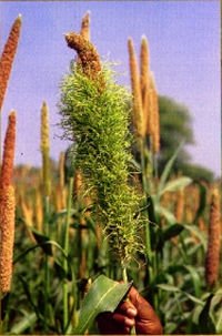
Crazy top downy mildew

Crazy top downy mildew (Sclerospora graminicola) on millet
(c) DFID Plant Sciences Research Programme (http://www.dfid-psp.org)

Crazy Top Downy Mildew

(Sclerospora graminicola)

Credits’: Biovision-Infonet

Crazy top downy mildew (Sclerospora graminicola) on millet
(c) DFID Plant Sciences Research Programme (http://www.dfid-psp.org)

Symptoms often vary as a result of systemic infection. Leaf symptoms begin as chlorosis at the base and successively higher leaves show progressively greater chlorosis.

On the lower leaf surface of infected leaves greyish white fungal growth may be observed. Severely infected plants are generally stunted and do not produce panicles.

Green ear symptoms result from transformation of floral parts into leafy structures. The disease is prevalent during rainy seasons.

What to do:

  • Plant resistant varieties, if available
  • Remove diseased plants from the field
  • Rotate with pulses

Long Smut

(Tolyposporium penicillariae)

Credits: Biovision-Infonet

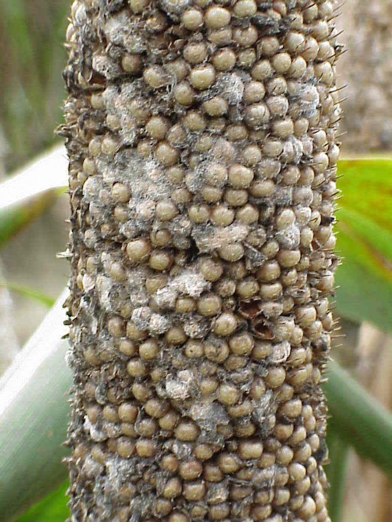
Long smut (Tolyposporium penicillariae)
(c) Kranz J., Schmutterer H., Koch W. Courtesy of EcoPort, www.ecoport.org

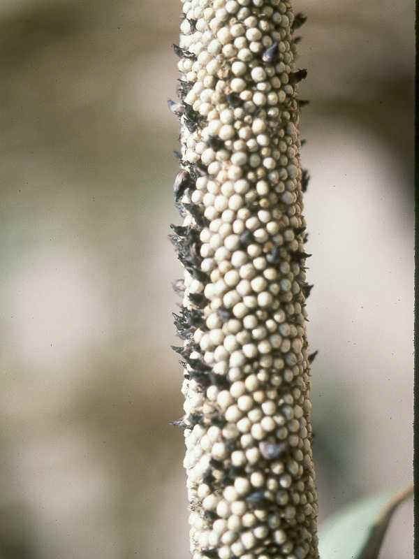
Immature, green fungal bodies (sori) larger than the seed develop on panicles during grain fill. A single fungal body (sorus) develops per floret. As grain matures, sori change in colour from green to dark brown. Sori are filled with dark spores.

Infection takes place at temperature range of between 21 and 31degC, and at relative humidity greater than 80%. The disease is spread by wind-borne spores and rain.

What to do:

  • Plant resistant varieties, if available.
  • Rotate with non-cereals.
  • Plough deep.
  • Practise good field sanitation.

Blast

(Pyricularia grisea)

Credits:Biovision-Infonet

Blast(Pyricularia grisea) on millet
(c) Reproduced from PEARL MILLET DISEASES – A Compilation of Information of the Known Pathogens of Pearl Millet (http://www.tifton.uga.edu/fat/pearlmilletdiseases.htm)

Lesions on foliage are elliptical or diamond-shaped, approximately 3 x 2 mm. Lesion centres are grey and water-soaked when fresh but turn brown upon drying.

Lesions are often surrounded by a chlorotic halo, which will turn necrotic giving the appearance of concentric rings. The disease is favoured by hot, humid conditions.

What to do:

  • Plant resistant varieties, if available.
  • Practise good field sanitation.

Ergot

(Claviceps spp.)

Credits: Biovision-Infonet

Ergot(Claviceps spp.) on millet
(c) Reproduced from PEARL MILLET DISEASES – A Compilation of Information of the Known Pathogens of Pearl Millet (http://www.tifton.uga.edu/fat/pearlmilletdiseases.htm)

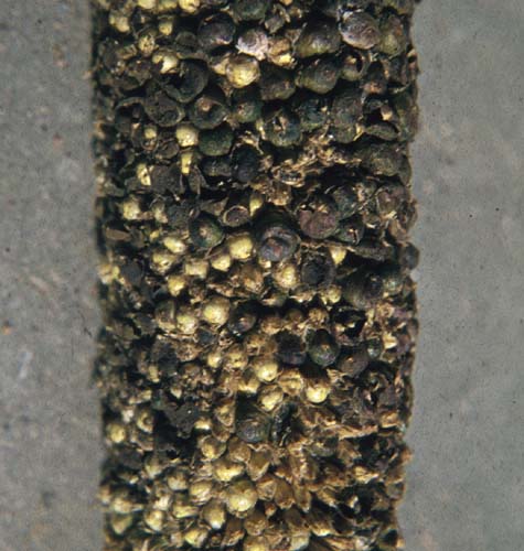
Cream to pink sticky “honeydew” droplets ooze out of infected florets on panicles. Within 10 to 15 days, the droplets dry and harden, and dark brown to black sclerotia (fungal fruiting bodies) develop in place of seeds on the panicle.

Sclerotia are larger than seed and irregularly shaped, and generally get mixed with the grain during threshing. Conditions favouring the disease are relative humidity greater than 80%, and temperatures between 20 to 30 degC.

The sclerotia falling on the soil or planted with the seed germinate when the plants are flowering. They produce spores that are wind-borne to the flowers, where they invade the young kernels and replace the kernels with fungal growth.

The fungal growth bears millions of tiny spores in a sticky, sweet, honeydew mass. These spores are carried by insects or splashed by rain to infect other kernels.

What to do:

  • Plant resistant varieties, where available.
  • Remove affected panicles.
  • Avoid planting seeds from infected panicles.
  • Plough deep.
  • Rotate with non-cereals preferably with pulses.
  • Practise good field sanitation.

Quick Search Millet Pests

Credits: Biovision-Infonet

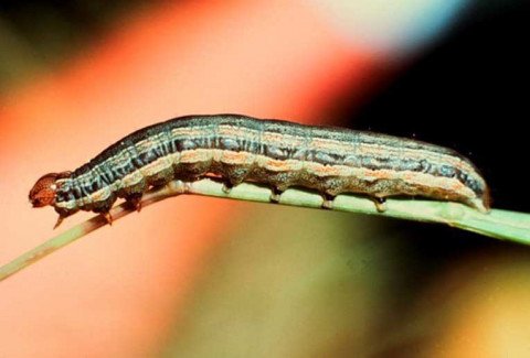
African armyworm

African armyworm (Spodotera exempta). Mature caterpillars measure up to 4 cm.
(c) University of Arkansas

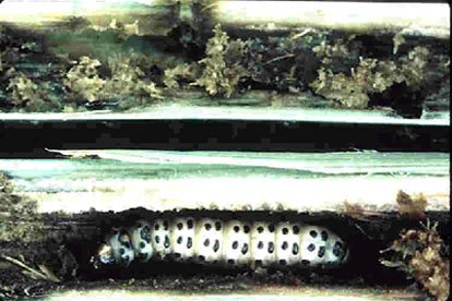
Stemborers

Active full-grown millet stemborer (Coniesta ignefusalis)
larva (feeding and damage) in millet stem.

(c) ICRISAT – International Crop Research Institute for the Semi-Arid Tropics. www.icrisat.org
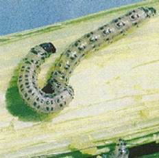
Spotted stemborer (Chilo partellus)
(c) Courtesy EcoPort (http://www.ecoport.org): Agricultural Research Council of South Africa.

Shoot fly

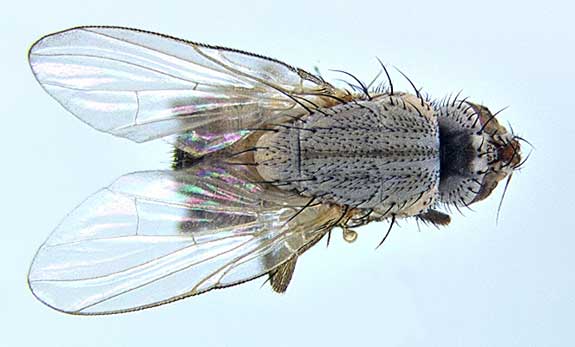
Shoot fly (Atherigona soccata) The adults are dark brown, and similar to a housefly, but nearly half the size (about 0.5 cm long).
(c) Georg Goergen, Courtesy of EcoPort, www.ecoport.org
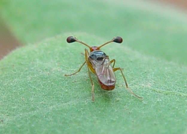
Stalk-eyed shoot fly (Diopsis spp.). It is about 8mm long.
(c) A.M. Varela, icipe

Grasshoppers

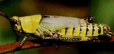
Variegated grasshopper (Zonocerus variegatus). The size of adult grasshoppers may vary between 3 – 5 cm
(c) PRIFAS. Courtesy of EcoPort, www.ecoport.org

Storage pests: The Lesser Grain Borer  and the Khapra beetle

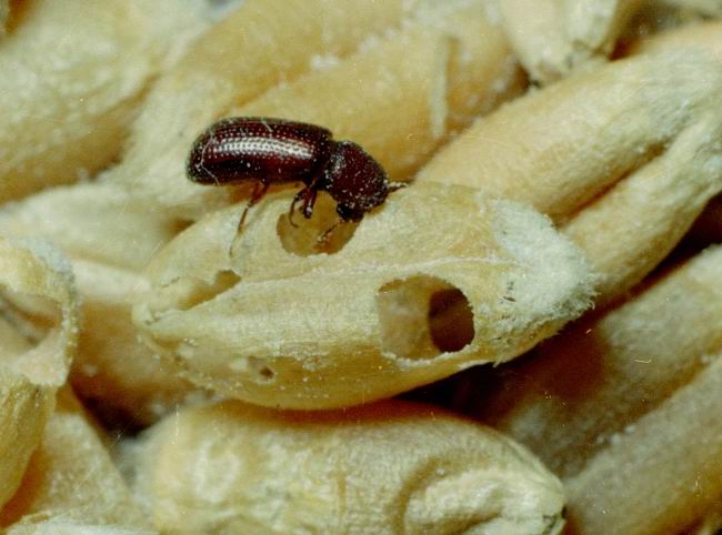
Lesser grain borer (Rhyzopertha dominica). Adults are 2-3 mm in length and reddish-brown in colour (shown on wheat grains).
(c) Clemson University – USDA Cooperative Extension Slide Series, United States, bugwood.org

Stemborers: African maize stalkborer

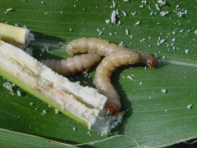
Caterpillars of the African maize stalkborer (Busseola fusca).
(c) D. Cugala, Stemborer team, icipe
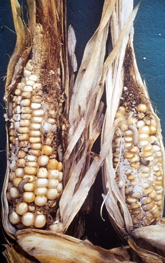
African maize stalkborer(Busseola fusca) damage on maize. Caterpillars are relatively featureless and noctuid, growing to a length of up to 4 cm. They lack conspicuous hairs or markings and look smooth and shiny. Colour is variable but usually creamy-white
(c) A. M. Varela, icipe

Mealybugs

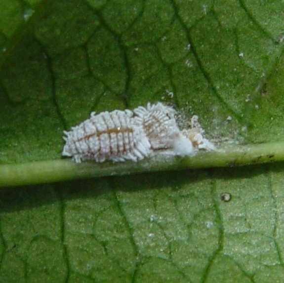
Female mealybugs on passionfruit leaf. Female mealybugs are 3 to 5 mm long and their body is usually covered with a waxy secretion.
(c) A.M. Varela, icipe